

What makes the human brain distinct?
Despite more than a century of study, we still don’t understand the root of our myriad mental abilities, such as abstract thought, language acquisition, and complex learning. We also don’t understand most brain diseases, some of which — like Alzheimer’s disease — seem to afflict only us out of all mammals.
Part of the difficulty in addressing these questions comes from the difficulty of studying the human brain at work. Many neuroscientists work with donated postmortem tissue, especially when studying individual brain cells.
An ongoing research program at the Allen Institute for Brain Science, a division of the Allen Institute, has found an unusual workaround to study live human neurons — they use pieces of live human brain removed during brain surgery and willingly donated to research. In the course of surgery for epilepsy or brain tumors, neurosurgeons often need to remove healthy pieces of tissue to access the diseased site. These pieces are transported to the laboratory for study, where neuroscientists are uncovering new information about human neurons while they are still alive.
Today, the Allen Institute research team and their collaborators have published an article in the journal Nature describing the details of those precious living cells donated by 90 patients, uncovering an increased diversity in certain types of human neurons as compared to mouse neurons. The study was part of a large, international collaboration brought together by the National Institutes of Health’s BRAIN Initiative to bring cutting-edge technologies to map the different types of brain cells in the human, monkey and mouse brains.
“The cell is, of course, the basic unit of life. Throughout the history of neuroscience, researchers have been trying to define the types of cells that make up the brain,” said Ed Lein, Ph.D., Senior Investigator at the Allen Institute for Brain Science, who led the study. “A fundamental part of what we’re trying to do is to create a census of cell types, which is similar to a census of people. We want to know what the whole population looks like, how many different types of cells are there are, and what their properties are.”
To build this census of the human brain, the scientists needed to get creative with new technologies. Their brain cell classification depends on a modern technology known as single-cell transcriptomics, which reads the complete set of genes a single cell switches on. Armed with this molecular method, the scientists homed in on cells in two layers of the 6-layered human neocortex, the wrinkled outermost shell of the brain and the seat of our higher cognitive functions.
The human neocortex is much larger compared to the rest of our brain than in other mammals, and the outer layers are especially expanded in primate evolution. The Allen Institute team found that humans have more types of neurons in these layers than mice do, and also uncovered new information about neurons that are especially susceptible to Alzheimer’s disease.
What makes the human brain different from a mouse brain?
There are many theories about how the human brain evolved to perform its modern cognitive capabilities, and there are likely many factors that drove that evolution. Do we have more of the same parts as other mammals, or did we evolve new, unique brain cells?
The answer, while far from complete, seems to be a bit of both.
One of the big surprises from the Human Genome Project, almost 20 years ago, was how similar our genes are to those of our closest ape relatives, and even to our not-so-close mammalian relatives. A comparable result emerges from looking at the cell types that make up the brain, which seem to be very similar to that of other mammals. However, there are key differences in our brain cell types, including specialization in specific kinds of cells, differences in the proportions of cell types and differences in the properties of cells.
The Allen Institute team decided to explore some of these key differences in the upper layers of the neocortex. Even at a glance, this area looks different in the mouse and human brain.
“In the human cortex, there’s really dense packing and small cell bodies in the upper part, and then as you go deeper, it gets sparser and you start to see giant cells. You don’t see that in the mouse. The corresponding layers in mouse cortex are just like one big block of dense small cells,” said Jonathan Ting, Ph.D., Assistant Investigator at the Allen Institute for Brain Science and one of the study’s co-first authors, along with the Allen Institute’s Jim Berg, Ph.D., Staci Sorensen, Ph.D., and Jeremy Miller, Ph.D. “That would make any curious scientist ask, what is driving that difference?”
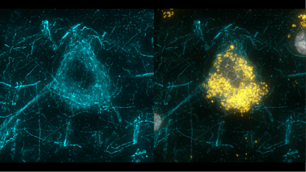
Using a technique called Patch-seq, which captures electrical activity, 3D shape, and gene expression all from the same individual neuron, the research team delved deep into hundreds of neurons in these layers in live tissue samples donated by surgery patients. The team, which included collaborating scientists who also study live human neurons from surgery patients at the University of Szeged in Hungary, Vrije Universiteit Amsterdam and The Hebrew University in Israel, focused on excitatory neurons, the kinds of neurons that activate other neurons.
They found that in these layers, humans have five types of excitatory neurons, as measured by Patch-seq, while mice have only three. Those two “extra” human neuron types are very obviously different from each other when you look at them, the researchers said. One kind, which the scientists dubbed CARM1P1 neurons for their most uniquely distinctive gene, are massive, highly elaborate cells with very long axons that seem to send long-range connections in the brain. The other, COL22A1 neurons, are smaller and scrawnier — Ting said they remind him of a stick figure drawing.
The increased diversity of neuron types in this region of the cortex is likely linked to our brain’s need for increased communication within the cortex, Lein said, an attribute that might underlie some of our enhanced mental abilities like abstract thought.
More than 30 years ago, neuroscientists found certain human neurons that make long-range connections are especially vulnerable in Alzheimer’s disease. That study also found an antibody that selectively marks these vulnerable cells. The Allen Institute team found that the largest cells in these layers, including the CARM1P1 neurons, seem to match those cells — they’re also marked with the specific antibody, and their 3D shape and gene expression match the disease-vulnerable cells found in the earlier study. The fact that these cells are lost in a disease that is marked by dramatic losses in memory and other cognitive functions suggests that the neurons could play an important role in human cognition. Uncovering their molecular identity could give scientists new pathways for better understanding — and treating — the cellular roots of Alzheimer’s disease.
“There’s strong evidence linking this molecularly defined cell population with a disease-vulnerable population that is responsible for long-range communication in the cortex,” Lein said. “This is a nice example of how what we call ‘cell typing’ can ultimately lead to a mechanistic understanding of what goes wrong in brains affected by dementia.”
The research described in this article was partially supported by several grant awards from institutes under the National Institutes of Health (NIH), including award U01MH114812 from National Institute of Mental Health, R01EY023173 from The National Eye Institute, and U01MH105982 from the National Institute of Mental Health and Eunice Kennedy Shriver National Institute of Child Health & Human Development, and R011EY023173 from The National Institute of Allergy and Infectious Disease. The content is solely the responsibility of the authors and does not necessarily represent the official views of NIH and its subsidiary institutes.
Citations
about the allen institute
The Allen Institute is an independent, 501(c)(3) nonprofit research organization founded by philanthropist and visionary, the late Paul G. Allen. The Allen Institute is dedicated to answering some of the biggest questions in bioscience and accelerating research worldwide. The Institute is a recognized leader in large-scale research with a commitment to an open science model. For more information, visit alleninstitute.org.


