in this article
A team of neuroscientists and engineers at the Allen Institute, Princeton University and Baylor College of Medicine has just released a collection of data that marries a 3D wiring diagram with the function of tens of thousands of neurons to create the most detailed examination of mammalian brain circuitry to date.
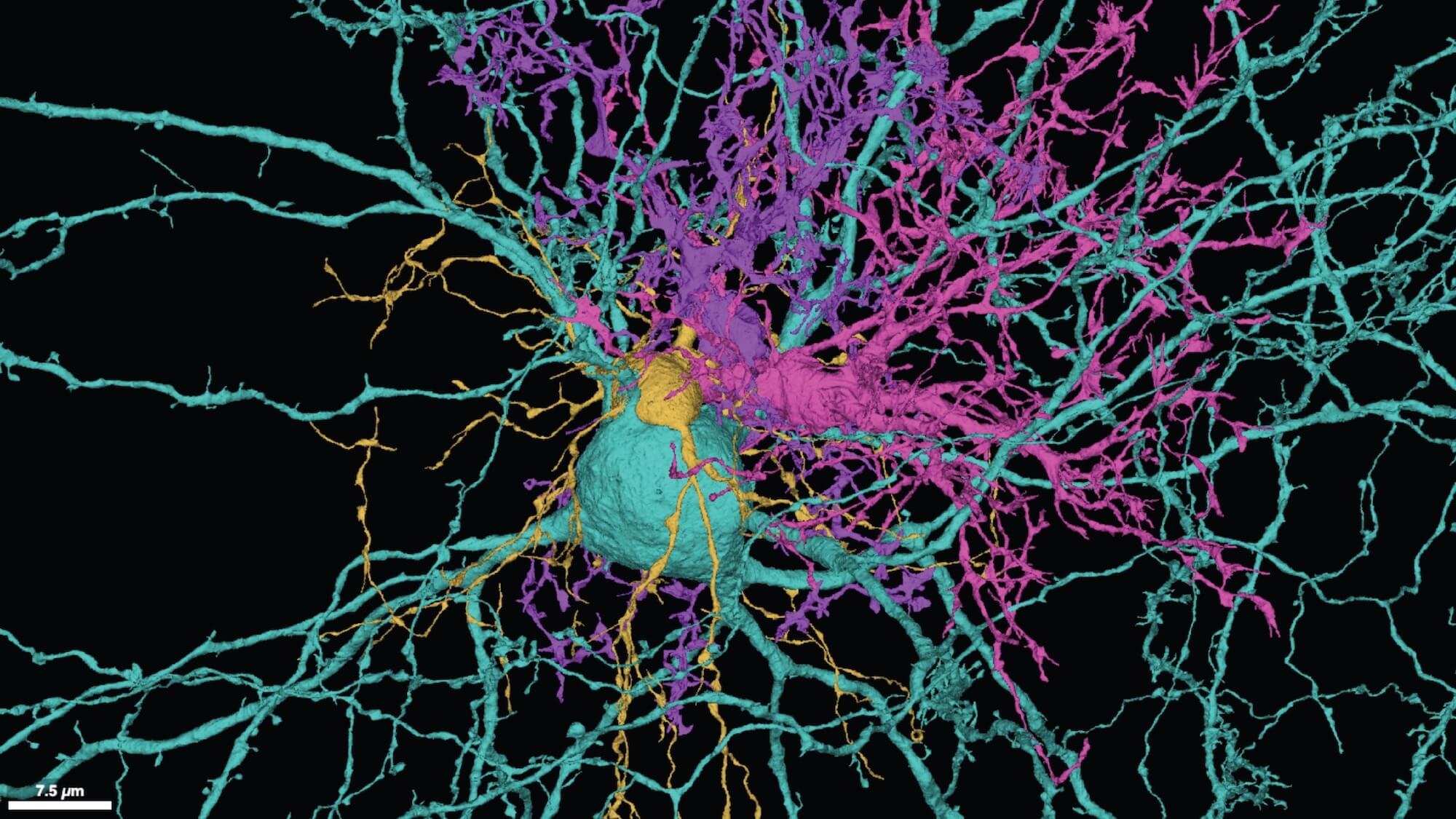
The dataset, which is publicly available for anyone in the community to browse and use, maps the fine structures and connectivity of 200,000 brain cells and close to 500 million synapses all contained in a cubic millimeter chunk of mouse brain — approximately the size of a grain of sand — from the visual neocortex, the part of the mammalian brain that processes what we see.
The public data collection is hosted online by the Brain Observatory Storage Service & Database (BossDB), and Amazon Web Services (AWS) is making the data freely accessible on the cloud through its Open Data Sponsorship Program. Google contributed storage and computing engine support through Google Cloud, and an open source visualization tool Neuroglancer through Google Research.
This massive project, dubbed the Machine Intelligence from Cortical Networks (MICrONS) program, took five years to complete and was funded and coordinated by the Intelligence Advanced Research Projects Activity, or IARPA. The goal of the research funding was to mine brain-wiring information to improve machine learning. But the dataset is also valuable to the neuroscience field — both for scientists seeking to understand how the brain conveys information along defined circuits, and for biomedical researchers wanting to treat brain disorders where wiring or connections are altered.

“We’re basically treating a brain circuit as a computer, and we asked three questions: What does it do? How is it wired up? What is the program?” said R. Clay Reid, M.D., Ph.D., Senior Investigator at the Allen Institute and one of the lead scientists of MICrONS. “Experiments were done to literally see the neurons’ activity, to watch them compute. The reconstructions that we’re presenting today let us see the elements of the neural circuit: the brain cells and the wiring, with the ability to follow the wires to map the connections between cells. The final step is to interpret this network, at which point we may be able to say we can read the brain’s program.”
The dataset joins the ranks of other recently released wiring diagrams from human and fruit fly brains captured using the same technology, electron microscopy or EM. This method reveals incredible details of an entire microscopic landscape — in this case, the densely packed topography of tendrilled neurons, astrocytes, blood vessels and other cells that make up the brain’s physical matter, as well as all the miniscule internal components of each cell.
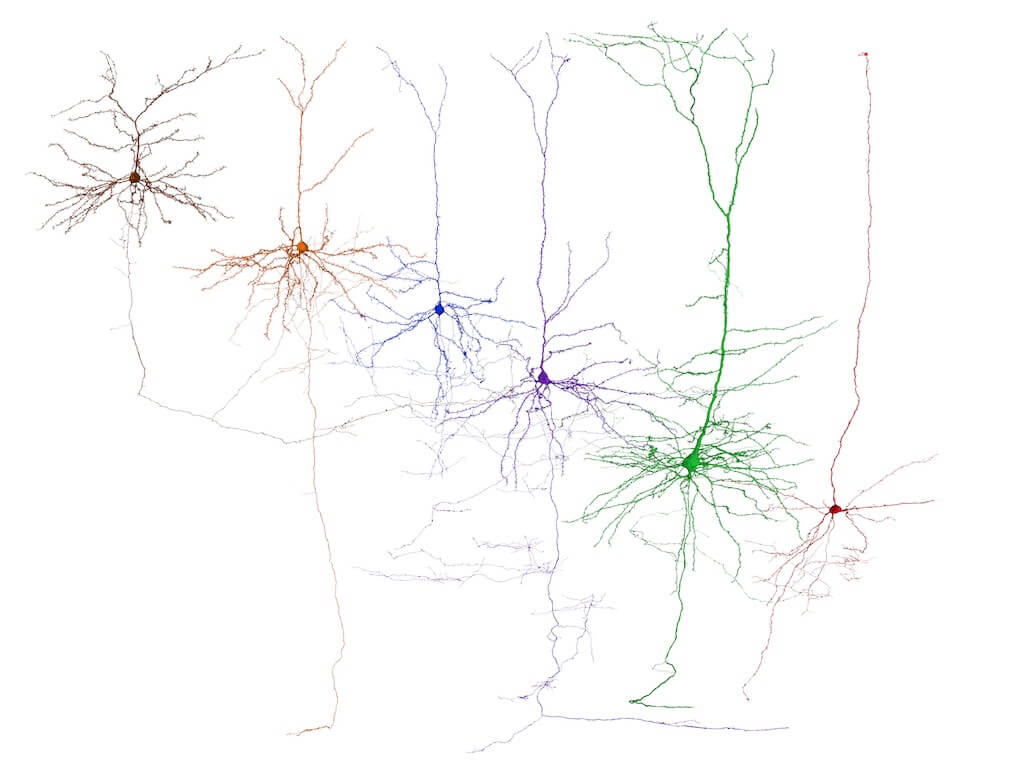
The newly released MICrONS data contains the most cells and connections of any such dataset to date, and it is large enough to capture entire local circuits and near-complete 3D shapes of individual mouse neurons. Some neurons make connections at incredibly long distances, sending their axons all the way across the brain, and those long-distance connections aren’t represented fully in this dataset. But the cubic millimeter volume was chosen to catch circuits across multiple brain areas involved in vision while also capturing the structure of as many entire neurons as possible. Before the structural data was gathered, the research team at Baylor captured neurons’ activity in this part of the brain as the mouse viewed images or movies of natural scenes.
“The neocortex contains billions of neurons communicating through trillions of connections that have endowed mammals with astonishing capabilities. A key question to untangling this bewildering complexity is to discover the relationships between the wiring rules and the functional properties of neurons,” said Andreas Tolias, Ph.D., Professor of Neuroscience and Director of the Center for Neuroscience and Artificial Intelligence at Baylor College of Medicine and one of the lead scientists of MICrONS. “This program is unique since it enabled us to assemble an interdisciplinary team to perform a very ambitious experiment that will enable us to answer this question.”
After the Baylor experiments, Allen Institute researchers preserved and sliced the brain piece in more than 27,000 equally thin slices, capturing a total of 150 million images of those slices using customized electron microscopes. The research team at Princeton then used machine learning approaches to “segment” the images, defining each cell and its internal components individually. The end result: beautiful, intricate digital renderings of 200,000 brain cells and the connections between them, many of which had never been captured in their complete form before.
“Our five-year mission had an ambitious goal that many regarded as unattainable. Today, we have been rewarded by breathtaking new vistas of the mammalian cortex,” said H. Sebastian Seung, Ph.D., Professor of Neuroscience and Computer Science at Princeton University and one of the lead scientists of MICrONS. “As we transition to a new phase of discovery, we are building a community of researchers to use the data in new ways. Only the collective efforts of this community can realize the resource’s potential for enabling new discoveries about the brain.”
Supported by the Intelligence Advanced Research Projects Activity (IARPA) via Department of Interior / Interior Business Center (DoI/IBC) under contracts D16PC00003 (Baylor), D16PC00004 (Allen), and D16PC0005 (Princeton). The U.S. Government is authorized to reproduce and distribute reprints for Governmental purposes notwithstanding any copyright annotation thereon. Disclaimer: The views and conclusions contained herein are those of the authors and should not be interpreted as necessarily representing the official policies or endorsements, either expressed or implied, of IARPA, DoI/IBC, or the U.S. Government.
Citations
about the allen institute
The Allen Institute is an independent, 501(c)(3) nonprofit research organization founded by philanthropist and visionary, the late Paul G. Allen. The Allen Institute is dedicated to answering some of the biggest questions in bioscience and accelerating research worldwide. The Institute is a recognized leader in large-scale research with a commitment to an open science model. For more information, visit alleninstitute.org.


