in this article
Meet cell 485105.03.02.01.
This cell started its life in a mouse brain, one of millions of other excitatory neurons, the broad class of cells whose job it is to activate other neurons. After months of research by multiple specialized teams, each investigating different attributes of the cell, its data — along with data from thousands of other mouse and human neurons — will help researchers around the world understand more about the brain.
Researchers in the electrophysiology lab search for specific brain cell types using fluorescent tags.
This bucket-brigade style of research is common in industry, but unusual among nonprofits. Most research organizations aren’t set up to conduct pipeline projects. But for large-scale datasets, conducting research via assembly line can be essential to generate high quality, standardized output, said Kimberly Smith, Associate Director of Molecular Biology at the Allen Institute for Brain Science, a division of the Allen Institute.
“If any one part of the chain, any one part of the team, doesn’t fulfill their duty to respect the standardization, the end product will suffer,” she said.
We decided to follow a single neuron through part of a pipeline, one that the researchers dub “Patch-seq.” Like other pipelines at the Allen Institute, this pipeline will result in open-access data for the broader scientific community. If all goes well, each neuron that enters the Patch-seq pipeline has the potential to yield information about its electrical behavior, 3D shape, and the set of thousands of genes it switches on.
Spoiler alert: cell 485105.03.02.01 won’t make it across all three of those finish lines.
How the neuron communicates
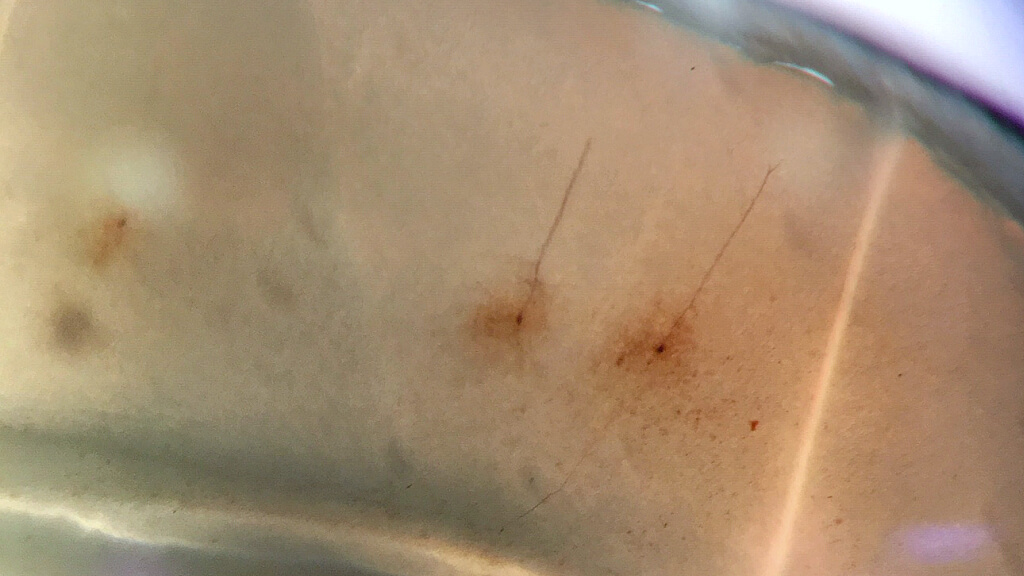
We joined cell 485105.03.02.01 on its naming day in the electrophysiology lab, the specialized laboratory where researchers, including research associate Jessica Trinh, probe uniquely identified neurons, still alive and firing inside a delicate slice of brain, with ultra-thin electrodes to eavesdrop on their electrical dispatches.
Trinh’s task is to record electrical signals from as many neurons as she can reliably get through. In a typical day, she records from 8 to 12. Once Trinh finds a healthy-looking cell among the many, she gently breaks its edge with a thin glass pipette – this is the “patch” part of Patch-seq. With a key stroke on the computer, her custom-built rig kicks into gear with a pre-programmed set of electrical tests. Red lines spike on the screen as cell 485105.03.02.01 reacts to the current coursing through the tiny electrode in its side. While she is recording the electrical signals, she is simulataneously filling the cell with a special dye that will later help another pipeline team to investigate its shape.
Trinh next removes cell 485105.03.02.01’s nucleus, the central storage compartment that holds all the neuron’s DNA. In one slow, smooth motion, she pulls out the clear sac, then deposits it in a tiny tube, to be frozen and sent to another team down the hall.
Staining the cell
Several days later, cell 485105.03.02.01 waits in the histology lab for the team that will prepare the brain slices for imaging. Their work involves a series of chemical reactions over several days that stain the slices for detailed microscopic photos. The histology team does many of their steps in pairs, one person acting as a witness to make sure no steps are skipped.
“As silly as it sounds, it keeps us accurate,” said Alice Pom, a research associate in the lab.
After staining, they check each slice under the microscope. The neuron’s body is dark brown on a pale tan background, its branching tendrils like lace in the sepia tones. They use tiny paintbrushes to ease the slice onto a glass microscope slide, and then stack several finished slides into boxes for the next team.
The cell’s genes
A week after its electrical debut, cell 485105.03.02.01’s nucleus waits, frozen, in a tiny tube in the molecular biology lab. Research associate Delissa McMillen starts her day early, preparing several hundred tubes at once for analysis. She adds chemical mixtures to the tubes to pulverize the nucleus’ membrane and prepare its RNA, the molecule that shows which genes the neuron has switched on when it was still alive, for further analysis.
McMillen came up with mental systems to make sure she’s doing all the steps in the correct order. She reads out numbers as she goes, opening and closing the tiny tubes, which are easy to drop if you’re not careful, she said.
“I always think about all the other researchers who worked for months to get the cells to this point, so I want to do my best to do my work the right way,” she said. “Every step takes trust. You have to trust that the next people are going to do their best with all the work you just put into their hands.”
Ready for its closeup
The following week, the slide containing the neuron’s body arrives in the imaging lab, where researchers take photos through powerful microscopes. Cell 485105.03.02.01’s first photoshoot is relatively quick, taken at a magnification of 20 times its actual size to provide context about the cell’s location in the brain.
Much of their work is automated, but the photos still require expertise and the human eye to pick out the delicate branching axons and dendrites from the background, making sure the camera is capturing what it should.
“It’s pretty cool to get to understand the full 3D essence of the cell,” said Shea Ransford, a research associate who works on the imaging team. “For some human neurons (which are much larger than mouse neurons), the projections just keep going; you can visibly track how far that cell has traveled in the brain.”
Once the cell passes a quality control check, the imaging team then takes a more detailed, more magnified photo, which can take upwards of five hours to complete for a particularly complex cell. The microscopes must stay completely still while imaging. One week, nearby construction jostled the cameras out of focus as they were running, setting the team’s work back by several days.
The go/no-go decision
Rachel Dalley, a senior research associate who leads a team of researchers who trace neurons’ precise 3D shapes, or their morphology, evaluates the first round of images of cell 485105.03.02.01. She points out to us where the cell’s “apical dendrite,” the long tendril that should reach from the cell’s body all the way to the outermost shell of the brain, disappears in the photo where it was cut off when the brain was sliced. Cell 485105.03.02.01 will not move on to the next stage in the pipeline, Dalley decides. Its experimental journey is done.
But its time in the lab was not in vain. There’s a lot scientists can learn from neurons’ electrical activity and gene expression alone. In fact, much of the data that will be released from the Patch-seq pipeline later this year will only include those two modalities. The cell’s shape is so delicate that, like cell 485105.03.02.01, many neurons in the pipeline don’t pass the quality control necessary to warrant the time-intensive, mostly manual reconstruction.
The cells’ shapes
Meet cell h48.06.368.11.14.01.02, a human neuron. We don’t know the details of this particular person — that information is kept anonymous — but we know that they underwent brain surgery in a Seattle hospital for either epilepsy or a tumor and donated a healthy piece of brain tissue removed during the course of surgery to research.
We caught up with this human neuron toward the very end of its pipeline journey, when it was already pixels on a computer monitor in the morphology lab. Research associate Grace Williams demonstrated the custom-made software that allows her to capture every detail of the neuron’s complex, branching axon and dendrites. It’s akin to a tricky, three-dimensional connect-the-dots diagram, Williams said.
Tracing the neuron’s morphology is a laborious process. A single complicated neuron can take up to three full days to reconstruct, Williams said, “but it is cool to think about how much work was already done before we get the image, and how hard that was.”
Williams and her colleague Alice Mukora described some of the tactics the team uses to deal with repetition, a common theme for many of the researchers involved in the pipeline. They pay a lot of attention to ergonomics to avoid injury from hours of (computer) mouse work. Mukora, who worked in a neuroscience lab in college before joining the Allen Institute, said that sometimes it helps to view her work through the lens of neuroanatomy: “What am I looking at in this cell? Does this structure make sense, from what I know about this particular kind of neuron?”
But sometimes it’s just about picking out the best podcasts to keep her focused through the next several hours of tracing.
The analysis
Once all the data is generated, it will be packaged and deposited on a public website for anyone to access. In the meantime, Allen Institute researchers are also plumbing the data to gain new insights into the types of cells that make up our brains. Computational neuroscientist Nathan Gouwens, Ph.D., uses methods he and his colleagues have developed to sort the data from the three branches of the pipeline into different buckets — and then studying how those buckets fit together — to give a measured view of brain cell type categories.
The researchers hypothesize that a brain cell type definition will be consistent, no matter if you are looking at its shape, electrical activity or its genes. But that remains to be proven — they’re still in the middle of sifting through the data from the more than 3,000 neurons that have come through the Patch-seq pipeline. This is the first time at the Allen Institute that these three kinds of data have been extracted from the same individual neurons across so many cells.
“It is kind of overwhelming being toward the end of such a large-scale effort,” Gouwens said. “But we are really trying to ensure we take advantage of this valuable dataset and learn as much as we can from these cells.”
Citations
about the allen institute
The Allen Institute is an independent, 501(c)(3) nonprofit research organization founded by philanthropist and visionary, the late Paul G. Allen. The Allen Institute is dedicated to answering some of the biggest questions in bioscience and accelerating research worldwide. The Institute is a recognized leader in large-scale research with a commitment to an open science model. For more information, visit alleninstitute.org.


