in this article
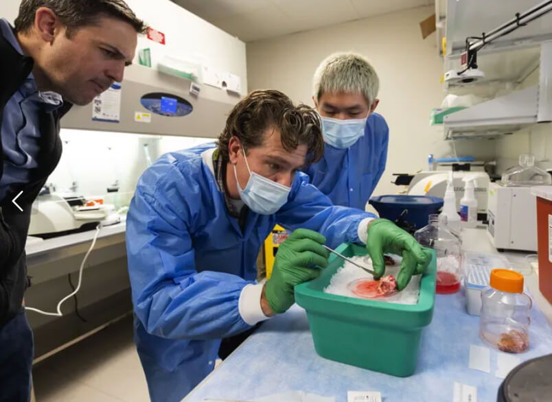
“You have to try to work as fast as you can, knowing you only have a few hours to extract the information you need,” said Ting, an assistant investigator at the Allen Institute for Brain Science.
That’s because mouse brain tissue generally survives for upwards of six hours in the lab. And if you’re ultimately interested in how the human brain works, as Ting is, such studies have another big drawback: Mouse brain cells are different from human brain cells in ways that researchers are only beginning to understand. That means these animals may not always be the best models to understand or treat human brain diseases, especially psychiatric disorders.
Ting and his colleagues were intrigued when anecdotal evidence began trickling out that human brain tissue had longer lasting power than rodent samples. These occasional hints, which were essentially hidden in the fine print of scientific articles, pointed to something potentially big for the field of neuroscience: the possibility for developing new strategies to directly label and manipulate living human brain cells in the lab.
But nobody had proven in a systematic way that those studies could be done on adult human brain tissue, Ting said. So in 2014, that’s what he and his colleagues set out to do.
Using small pieces of brain removed during surgery by the team’s neurosurgeon partners at the Swedish Neuroscience Institute and Harborview Medical Center, Ting and other researchers at the Allen Institute for Brain Science have now shown that human brain tissue, provided a steady supply of oxygen, can survive for three days on a laboratory benchtop.

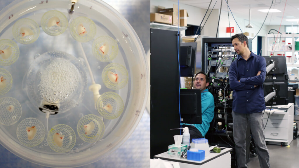
Viral labeling of live human neurons
The fact that human brain tissue can survive for days in the lab means neuroscientists can label and manipulate these precious cells to study them directly. Here, engineered human neurons flicker green when they are triggered to fire using a special neuron-activating chemical known as carbachol.
They also found that those precious few days are long enough to apply cutting-edge research techniques, originally developed for use in animal models, to perform experiments on living human brain tissue. In an initial set of experiments, the research team used a modified virus to insert designer genes in the human neurons that causes them to glow green under the microscope when they are active – and then watched them firing in real time when they were switched on by a neuron-activating chemical.
This rapid viral labeling approach will open up new research avenues for neuroscientists who want to study the human brain, especially at the level of individual cells, said Ting, who is a lead author on the study describing human brain tissue’s “exceptional viability,” published last week in the journal Scientific Reports. The researchers believe this discovery opens doors not only to understanding more about how the healthy human brain works, but to building a platform to better study brain diseases like epilepsy – or even to test the efficacy of potential new gene therapies before they’re ready for clinical trials.
That’s an exciting possibility for a field that’s seen so many animal studies fail to translate to human benefit.
“One thing we’re learning from studying human tissue is that there are a lot of differences between the rodent brain and the human brain,” said Brian Kalmbach, Ph.D., a neuroscientist at the Allen Institute for Brain Scientist and author on the study. “For translational research, the implications of that are pretty profound.”
If a researcher finds that a specific type of neuron seems to be defective in a mouse model of Alzheimer’s disease, for example, there’s no guarantee that the same mechanisms are at play in human disease. “The mouse is a good model system but identifying the differences from humans is important,” Kalmbach said. This new system will allow researchers to delve into those differences, cell by cell.
Therapeutic potential of the finding
Ryder Gwinn, M.D., a neurosurgeon at Swedish Neuroscience Institute who provided pieces of brain tissue from his epilepsy patients who underwent surgery, can see obvious applications of the finding for the field of epilepsy research. There’s a longstanding theory that epileptic seizures destroy a certain type of neuron that can suppress the activity that causes the seizures themselves. Over time, the theory goes, the condition gets worse because more and more of these inhibitory neurons die off, meaning seizures occur more easily and more often. But there’s no convincing proof of this theory yet, said Gwinn, who is also an author on the study.
With the advent of a system to study and genetically manipulate human brain tissue in the lab, that hypothesis could now be tested directly using tissue removed from epilepsy patients. And eventually, the system could also be used to test gene therapies that would enhance inhibitory signals, Gwinn said, potentially combatting epilepsy at its root.
“It’s the therapeutic implications of these findings that are really the most exciting from a clinician’s perspective,” Gwinn said. “This is a much more realistic way to test the viability of genetic therapies in humans than in a rodent model.”
Although the type of modified virus Ting and his colleagues used in the current study isn’t typically used for human gene therapies, it’s a short step from there to testing more clinically relevant tools, he said. The team is now using a different type of virus in their studies with similar success, one that is already being tested in human gene therapies for other applications.
Along with the downstream implications of their work for medical research, the researchers are inspired by the patients who are facing major surgeries for either epilepsy or brain tumors who agree to donate their brain tissue for research, Ting said.
“Basic research is probably the furthest thing from these patient’s minds when they are preparing to go under the knife for brain surgery, but almost everybody consents to donate tissue,” Ting said. “It’s a really remarkable view of their humanity and altruism, that this research could help us better understand their diseases down the road to come up with better treatments for other people.”
Citations
about the allen institute
The Allen Institute is an independent, 501(c)(3) nonprofit research organization founded by philanthropist and visionary, the late Paul G. Allen. The Allen Institute is dedicated to answering some of the biggest questions in bioscience and accelerating research worldwide. The Institute is a recognized leader in large-scale research with a commitment to an open science model. For more information, visit alleninstitute.org.


