in this article
From a tiny sample of tissue no larger than a grain of sand, scientists have come within reach of a goal once thought unattainable: building a complete functional wiring diagram of a portion of the brain. In 1979, famed molecular biologist, Francis Crick, stated that it would be “[impossible] to create an exact wiring diagram for a cubic millimeter of brain tissue and the way all its neurons are firing.” But during the last seven years, a global team of more than 150 neuroscientists and researchers has brought that closer to reality.
Scientists have created the largest wiring diagram and functional map of an animal brain to date.
The Machine Intelligence from Cortical Networks (MICrONS) Project has built the most detailed wiring diagram of a mammalian brain to date. Today, scientists published the scientific findings from this massive data resource in a collection of ten studies in the Nature family of journals. The wiring diagram and its data, freely available through the MICrONS Explorer, are 1.6 petabytes in size (equivalent to 22 years of non-stop HD video), and offer never-before-seen insight into brain function and organization of the visual system.
“The MICrONS advances published in this special issue of Nature are a watershed moment for neuroscience, comparable to the Human Genome Project in their transformative potential,” said David A. Markowitz, Ph.D., former IARPA program manager who coordinated this work.
“IARPA’s moonshot investment in the MICrONS program has shattered previous technological limitations, creating the first platform to study the relationship between neural structure and function at scales necessary to understand intelligence. This achievement validates our focused research approach and sets the stage for future scaling to the whole brain level.”

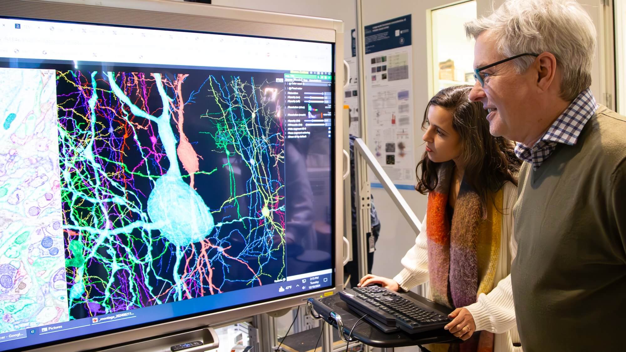
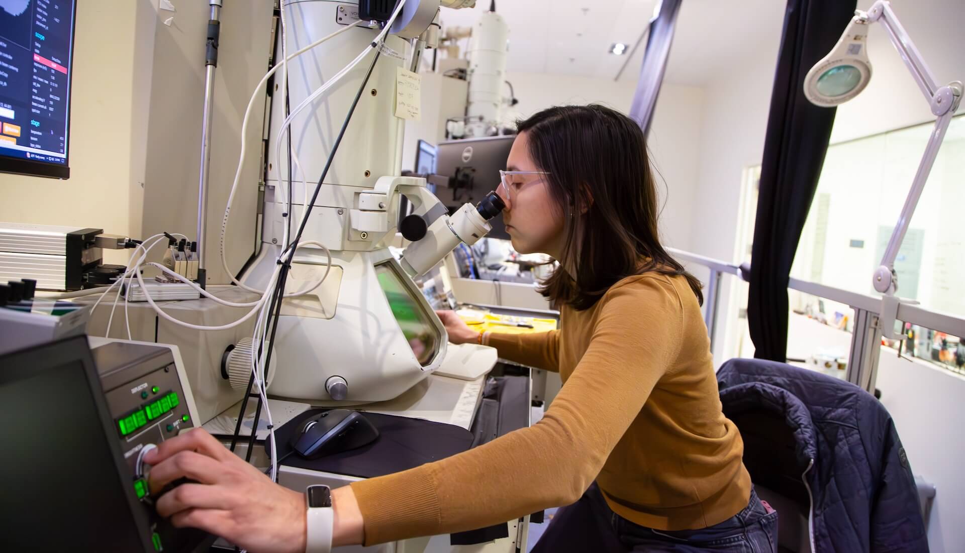
Scientists at Baylor College of Medicine and Stanford University began by using specialized microscopes to record the brain activity from a one cubic millimeter portion of a mouse’s visual cortex as the animal watched various movies and YouTube clips. Afterwards, Allen Institute researchers took that same cubic millimeter of the brain and sliced it into more than 25,000 layers each 1/400th the width of a human hair and used an array of electron microscopes to take high-resolution pictures of each slice. Finally, another team at Princeton University used artificial intelligence and machine learning to reconstruct the cells and connection into a 3D volume. Combined with the recordings of brain activity, the result is the largest wiring diagram and functional map of the brain to date, containing more than 200,000 cells, four kilometers of axons – the branches that reach out to other cells, and 523 million synapses – the connection points between cells.
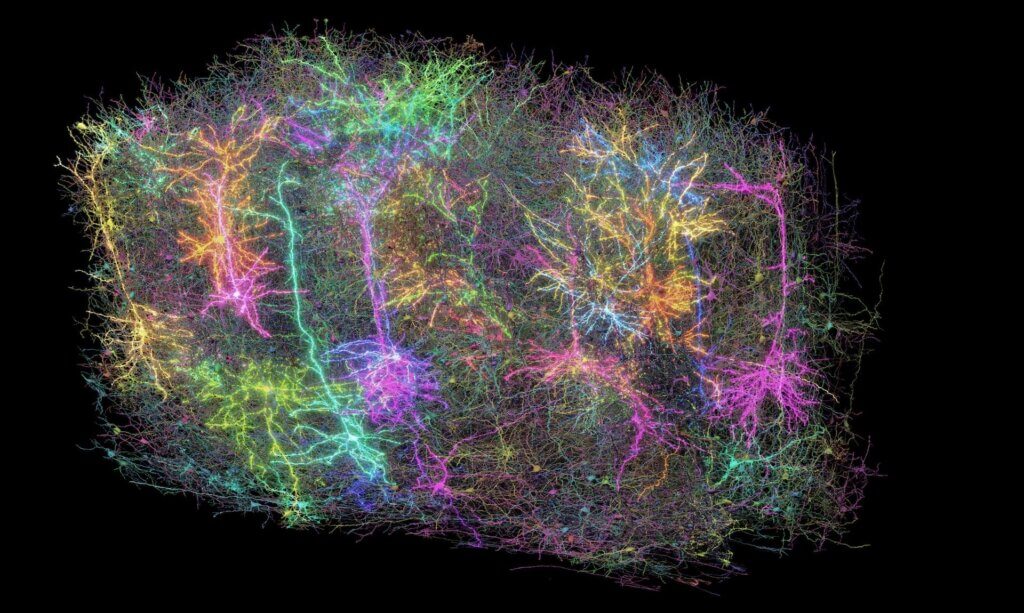
“Inside that tiny speck is an entire architecture like an exquisite forest,” said Clay Reid, M.D., Ph.D., senior investigator and one of the early founders of electron microscopy connectomics who brought this area of science to the Allen Institute 13 years ago. “It has all sorts of rules of connections that we knew from various parts of neuroscience, and within the reconstruction itself, we can test the old theories and hope to find new things that no one has ever seen before.”
A New Look at Brain Function and Organization
The findings from the studies reveal new cell types, characteristics, organizational and functional principles, and a new way to classify cells. Among the most surprising findings was the discovery of a new principle of inhibition within the brain. Scientists previously thought of inhibitory cells—those that suppress neural activity—as a simple force that dampens the action of other cells. However, researchers discovered a far more sophisticated level of communication: Inhibitory cells are not random in their actions; instead, they are highly selective about which excitatory cells they target, creating a network-wide system of coordination and cooperation. Some inhibitory cells work together, suppressing multiple excitatory cells, while others are more precise, targeting only specific types.

“This is the future in many ways,” explained Andreas Tolias, Ph.D., one of the lead scientists who worked on this project at both Baylor College of Medicine and Stanford University. “MICrONS will stand as a landmark where we build brain foundation models that span many levels of analysis, beginning from the behavioral level to the representational level of neural activity and even to the molecular level.”
Scientists present the largest, most detailed, functional wiring diagram of a mammalian brain to date
What this Means for Science and Medicine
Understanding the brain’s form and function and the ability to analyze the detailed connections between neurons at an unprecedented scale opens new possibilities for studying the brain and intelligence. It also has implications for disorders like Alzheimer’s, Parkinson’s, autism, and schizophrenia involving disruptions in neural communication.
“If you have a broken radio and you have the circuit diagram, you’ll be in a better position to fix it.” said Nuno da Costa, Ph.D., associate investigator at the Allen Institute. “We are describing a kind of Google map or blueprint of this grain of sand. In the future, we can use this to compare the brain wiring in a healthy mouse to the brain wiring in a model of disease.”
Collaboration Across Borders

The MICrONS Project is a collaborative effort of more than 150 scientists and researchers from the Allen Institute, Princeton, Harvard, Baylor College of Medicine, Stanford and many others.
“Doing this kind of large, team-scale science requires a lot of cooperation,” said Forrest Collman, Ph.D., associate director of data and technology at the Allen Institute. “It requires people to dream big and to agree to tackle problems that aren’t obviously solvable, and that’s how advances happen.”
The collaborative, global effort was made possible by support from the Intelligence Advanced Research Projects Activity (IARPA) and National Institutes of Health’s Brain Research Through Advancing Innovative Neurotechnologies® Initiative, or The BRAIN Initiative®.
“The BRAIN Initiative plays a critical role in bringing together scientists from various disciplines to perform complex and challenging research that cannot be achieved in isolation,” said John Ngai, Ph.D., director of The BRAIN Initiative®. “Basic science building blocks, like how the brain is wired, are the foundation we need to better understand brain injury and disease, to bring treatments and cures closer to clinical use.”
A map of neuronal connectivity, form, and function from a grain of sand-sized portion of the brain is not just a scientific marvel, but a step toward understanding the elusive origins of thought, emotion, and consciousness. The “impossible” task first envisioned by Francis Crick in 1979 is now one step closer to reality.
Citations
about the allen institute
The Allen Institute is an independent, 501(c)(3) nonprofit research organization founded by philanthropist and visionary, the late Paul G. Allen. The Allen Institute is dedicated to answering some of the biggest questions in bioscience and accelerating research worldwide. The Institute is a recognized leader in large-scale research with a commitment to an open science model. For more information, visit alleninstitute.org.


