
Brian Kalmbach, Ph.D., was on a hunt for a very specific part of a very specific cell.
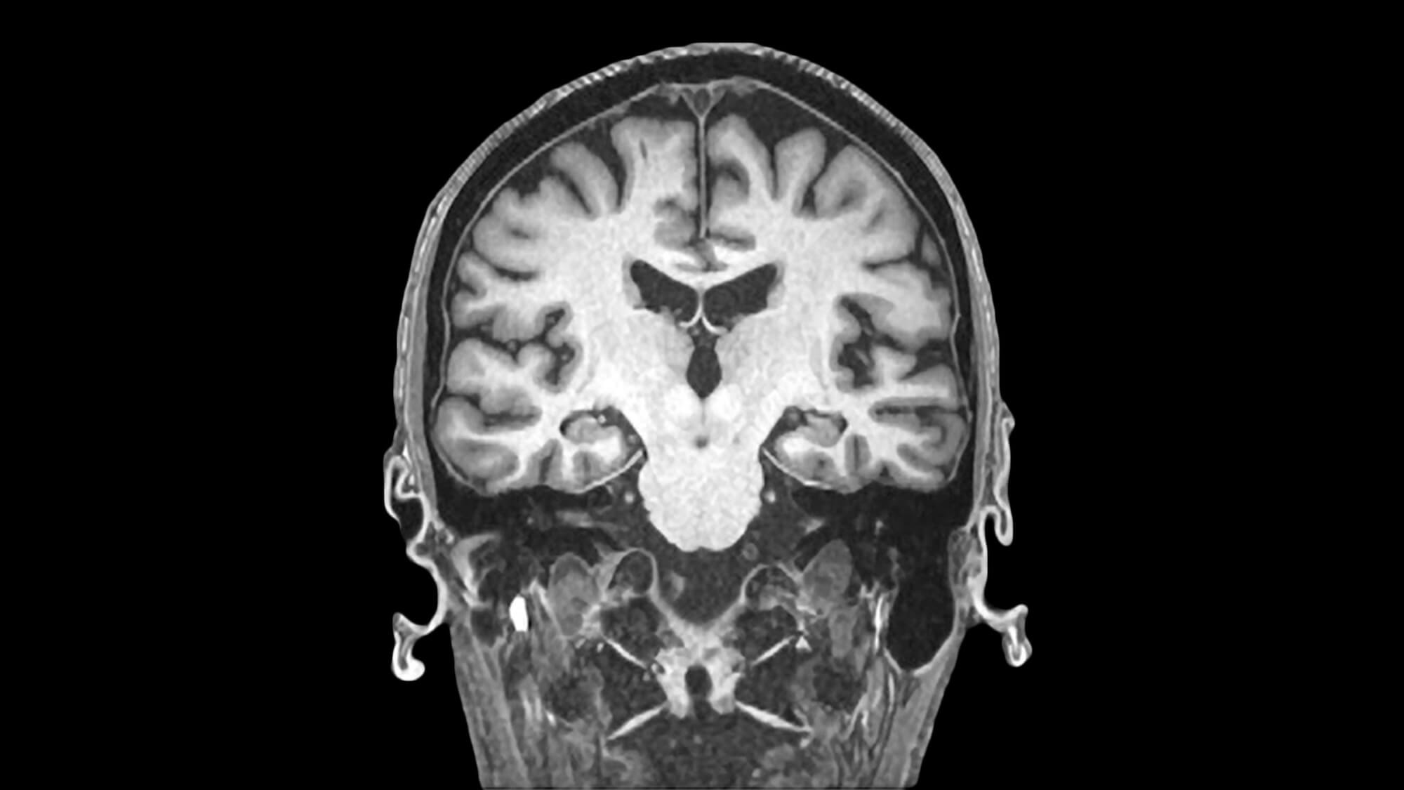
It wasn’t an easy hunt. No fun scavenger clues on the way. This human brain cell, still alive, was buried near the very bottom of a thin slice of the many-layered cortex, the wrinkled outermost shell of the brain. The fifth layer of the cortex, where the scientist’s target lived, is chock-full of myelin, the tough white support material that surrounds nerve bundles.
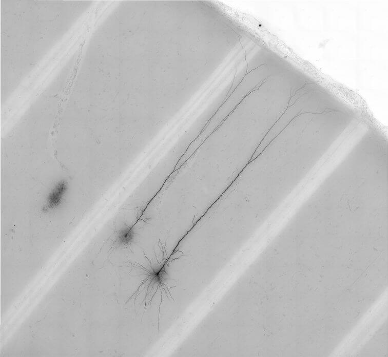
And Kalmbach was aiming not for the largest part of the neuron, its soma or cell body, but for its dendrites, the skinny tendrils that branch in many directions from one end of the soma. Even with the help of a powerful microscope, the dendrites are translucent and difficult to pick out from the background.
“You really have to train your eyes to find them,” Kalmbach said. “Sometimes I feel like I’m chasing a mirage.”
That specific neuron was one of just a few dozen of its kind that Kalmbach and his colleagues at the Allen Institute for Brain Science, a division of the Allen Institute, studied in detail from living human brain tissue. The rare human neurons the team hunted down in these donated tissue samples are known by a somewhat complex name: layer 5 extratelencephalic-projecting neurons. The scientists call them L5 ET neurons for short.
The Allen Institute team and their collaborators published a study in the journal Neuron today describing the electrical properties, gene expression and 3D shapes of human L5 ET neurons, the first such study to capture all these features of these rare human brain cells. And they also made new discoveries about how the cells produce distinctive electrical signals from the tiny dendrites that are so difficult to find in a mass of gray and white.
L5 ET neurons are a class of excitatory neurons, the kinds of neurons that activate other neurons. Allen Institute scientists and other researchers have seen that human L5 ET neurons seem to have more specialized features as compared to their mouse counterpart. The massive von Economo neuron, a specialized kind of neuron that exists in humans, dolphins, cows and other large mammals but not in rodents, is a subtype of L5 ET neuron. Betz cells also fall into this class; these large motor neurons send their axons all the way to the spinal cord and degenerate in ALS, a devastating neurological disease that affects movements.
Kalmbach specializes in a type of neuroscience known as electrophysiology, which entails contacting the outer membrane of a neuron with a tiny electrode and goosing the neuron with current to “fire” action potentials, the neuron’s method of electrical communication. Usually, electrophysiologists take readings at the cell soma — textbook neuroscience tells us that action potentials start in the soma and travel down the axon, the neuron’s “transmitter.” For a long time, dendrites were thought to be passive receivers of electrical signals
Rodent L5 ET neurons are well-known for a different kind of signal — their dendrites can also generate spikes. In mice, researchers have found that this kind of signal is linked to conscious perception, and that when animals are under general anesthesia, the connection between dendritic signals and the rest of the neuron is severed.
It wasn’t clear whether dendritic spikes happened in human L5 ET neurons, which are much larger than their rodent counterparts; other researchers did not find these kinds of spikes in living human neurons. But when Kalmbach probed the L5 ET neurons’ dendrites, he saw signals of dendritic spikes that were clear as day.
“It’s really exhilarating to patch a human dendrite and see these spikes for the first time,” he said. “There are rare times as a researcher when you are doing a technically difficult experiment and you realize that you are seeing something that no one else in the world has seen.”
Rare but important
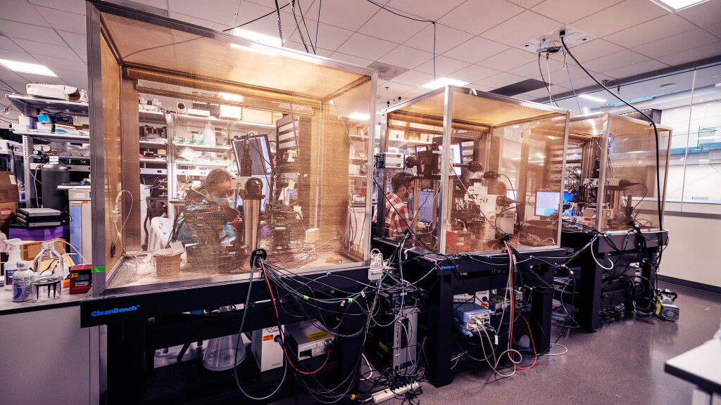
To study live human neurons, the Allen Institute team relies on donations from Seattle-area brain surgery patients. Healthy brain tissue is removed during surgery for epilepsy or tumors and, with the patients’ consent, is transported to the Allen Institute while it’s still alive for study. For the L5 ET neuron study, the researchers looked at brain tissue taken from the middle temporal gyrus, a section of the temporal lobe of the cortex that’s thought to be involved with visual tasks like perception of distance, facial recognition, and recalling word meaning during reading. This region of the cortex only contains one subtype of L5 ET neurons.
Because L5 ET neurons are defined by long projections that are entangled elsewhere in the brain and body, it’s even harder to know which neurons in these small pieces of brain were ET neurons and which are related but closer-projecting types. Here, the team turned to the cells’ genes. Using a technique known as Patch-seq, which captures a single cell’s electrical properties, 3D shape, and the suite of genes each cell switches on (also known as its transcriptome), the researchers were able to pinpoint human L5 ET neurons by comparing their transcriptomic profiles to those of laboratory mouse neurons.
Their dendritic spikes weren’t the only interesting things about these cells. The team found that compared to those of mice, human L5 ET neurons are much rarer in the cortex. The cells are a kind of excitatory neuron, neurons that activate other neurons. In mice, L5 ET neurons comprise up to 30% of excitatory neurons in layer 5; in human brains, they’re only about 5%. Given that L5 ET neurons hold so many specialized subtypes, like the von Economo neuron and the Betz cell, it’s intriguing that they also seem to be present in lower proportions in the human brain, the scientists said.
“These cells must be doing something really important, since they are rare but persisted during evolution,” said Jonathan Ting, Ph.D., Assistant Investigator at the Allen Institute for Brain Science and senior author on the study.
Although it’s not yet clear what role these rare neurons play in the human brain, the fact that they can generate dendritic spikes like their rodent counterparts suggests that L5 ET neurons might be involved in our own conscious perception as well. The research described in this article was supported in part by the National Institutes of Health awards U01 Mh414812-02, RF1Mh414126, RF1Mh421274, R01DA036909, AG005136, P51OD010425 and UL1TR000423. Its contents are solely the responsibility of the authors and do not necessarily represent the official views of the NIH.
Rachel Tompa is Senior Writer at the Allen Institute. She covers news from all scientific divisions at the Institute. Get in touch at rachelt@alleninstitute.org.
Citations
about the allen institute
The Allen Institute is an independent, 501(c)(3) nonprofit research organization founded by philanthropist and visionary, the late Paul G. Allen. The Allen Institute is dedicated to answering some of the biggest questions in bioscience and accelerating research worldwide. The Institute is a recognized leader in large-scale research with a commitment to an open science model. For more information, visit alleninstitute.org.


