
In the era of single-cell biology, genes are king.
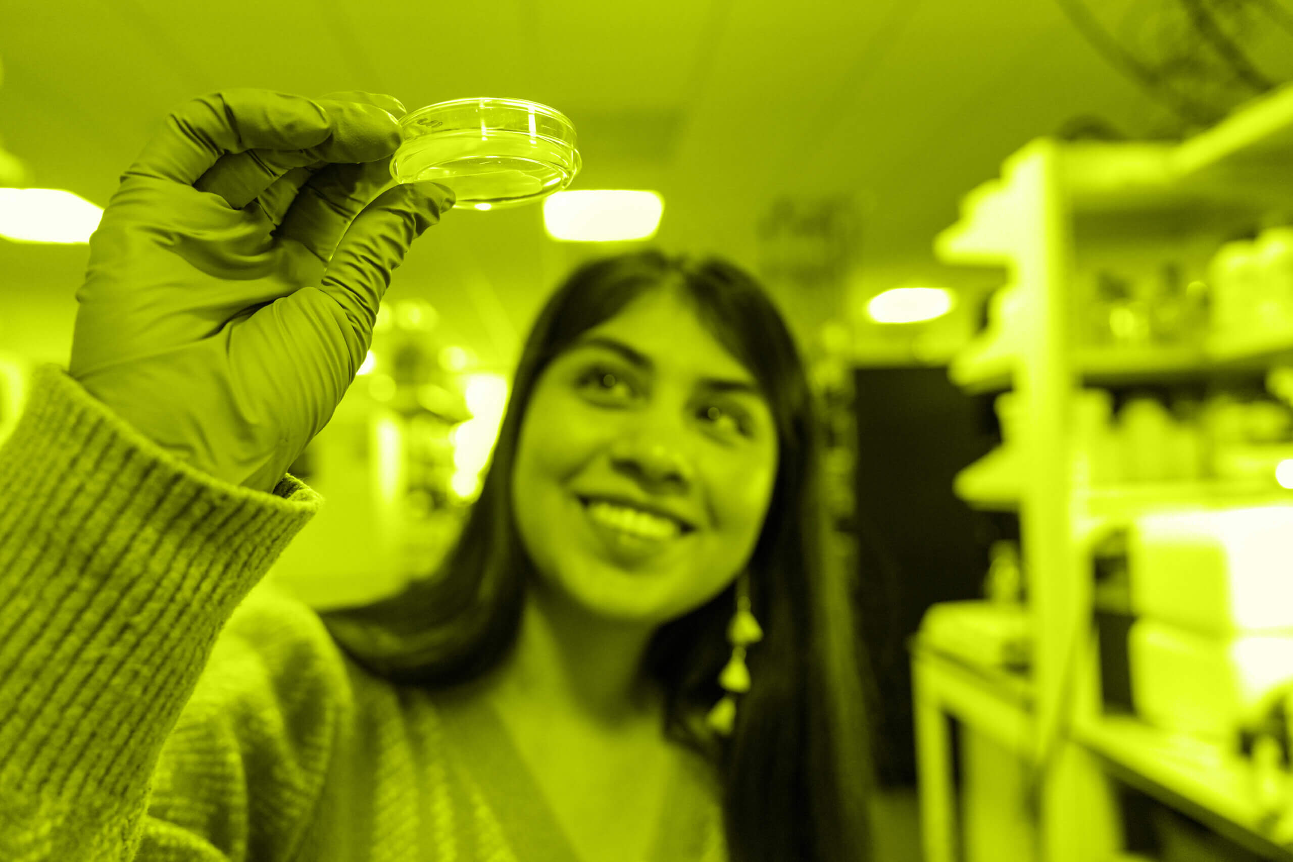
Single-cell research — which has exploded in popularity and feasibility in recent years — entails looking at some attribute of living things (cells in a petri dish, tissues, even entire organs) one cell at a time, and lots of them.
Initially, genes and their activity were arguably the easiest attribute to study using this approach. Scientists can now measure how many genes are switched on and by how much — the gene’s expression levels — in a quantitative way, relatively quickly, across all genes in an individual cell and across many hundreds of thousands or even millions of different cells. While other types of single-cell research are increasingly common, single-cell gene expression is still a go-to in many cell biology studies.
But a new study hints that if you want to really understand what a cell is doing, single-cell gene expression might not get you that far.
Researchers at the Allen Institute for Cell Science, a division of the Allen Institute, developed a method to automatically capture visual characteristics from thousands of images of human heart muscle cells generated from human stem cells. When they compared one important characteristic of those heart cells — a score for how “organized” the muscle cells’ contracting machinery was, a measure of the cells’ progress on the path to maturity — to expression of the genes responsible for heart cell maturity, they found very little correlation. The team published a study describing this method and their findings in the journal Cell Systems today; the study was also featured on the cover image of the journal.
“This paints a broader picture of our cells. If someone wants to really understand and characterize a cell’s state, we found that having both of these types of information can be complementary and tell you something different and unique about what’s going on in the cell. Basically, it’s a really complicated picture,” said Kaytlyn Gerbin, Ph.D., a scientist at the Allen Institute for Cell Science and one of four co-first authors on the Cell Systems publication, along with bioinformatics associate Tanya Grancharova, former Allen Institute for Cell Science scientist Melissa Hendershott, Ph.D., and former senior scientist Rory Donovan-Maiye, Ph.D.
How do you measure a picture?
To compare gene expression to cell structures, the scientists needed a way to translate images of cells to a quantitative measurement. While microscopy allows researchers to study individual cells, it’s usually not done at the same large scale as other types of single-cell research, and it’s not exactly straightforward to convert an image of cells to a number.
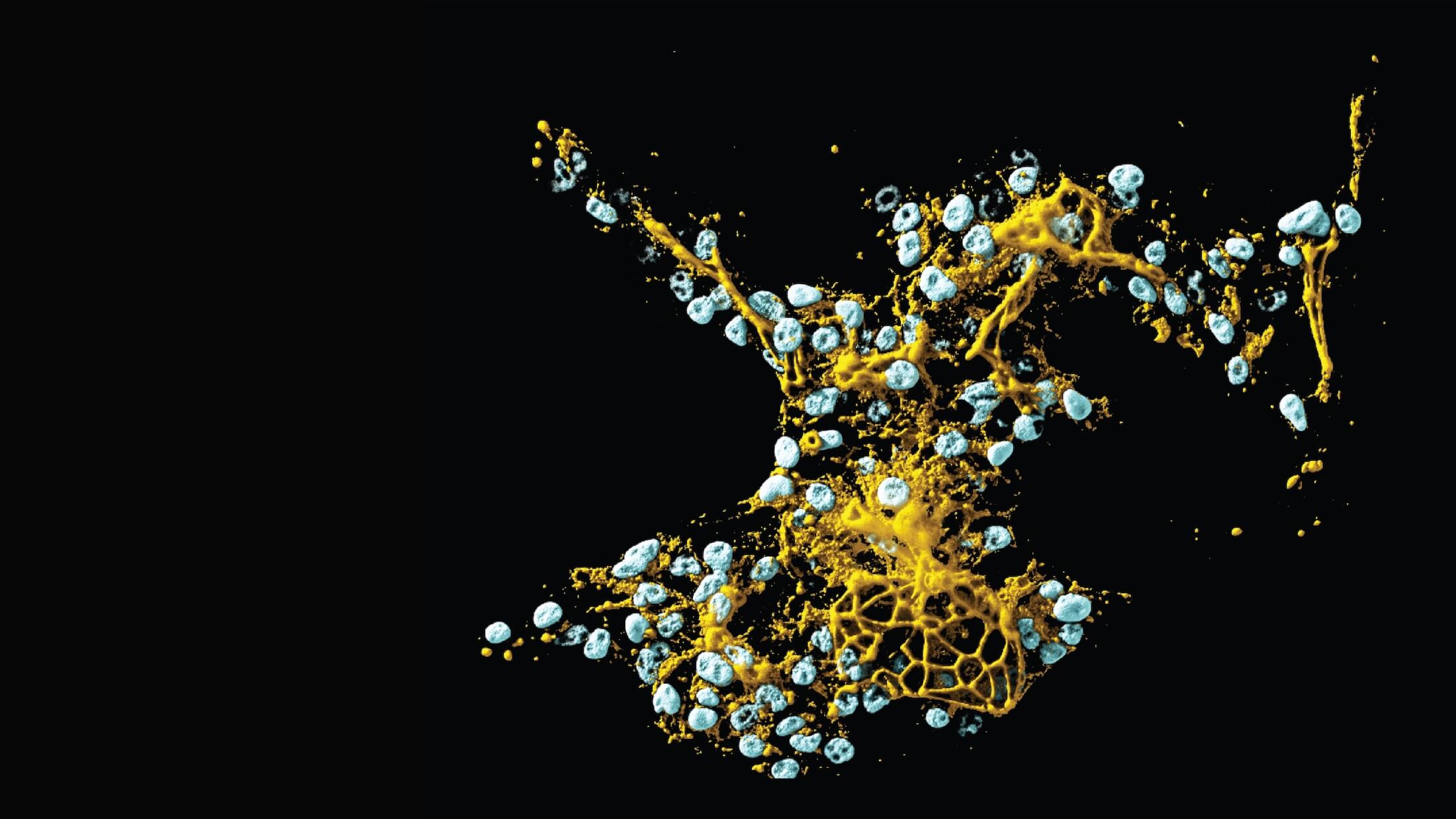
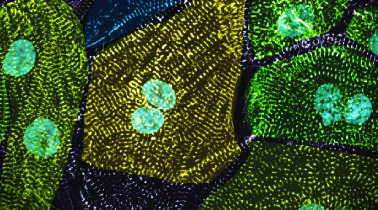
The researchers focused on a key structure in heart muscle cells known as the sarcomere. These tiny cellular structures allow the heart muscle cells, also called cardiomyocytes, to contract — they’re the driving force behind your heartbeat.
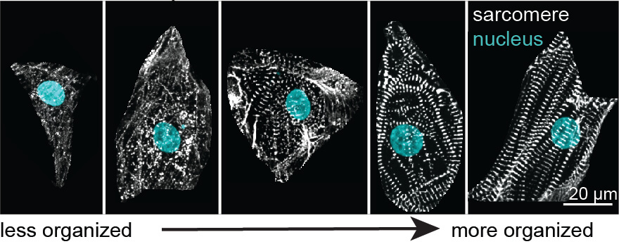
As naïve stem cells develop into cardiomyocytes, sarcomeres form and become more organized as heart cells mature. When sarcomeres first appear as stem cells start to turn into heart cells, they look like scattered dots, but they later elongate and line up perpendicular to the length of the long, skinny cells, like slats on a tiny railroad track.
The stripe-pattern structures contracting and expanding together cause cardiomyocytes to contract and expand in sync, allowing your heart to beat and pump blood around your body. Several genetic heart diseases are linked to poorly formed or unorganized sarcomeres; if the structures don’t fall into line as they should, the heart can’t beat correctly.
Heart cell maturation is also linked to increased expression of certain genes involved in sarcomere formation. One of these genes produces a protein called myosin, which is one of the key building blocks of the sarcomere. But nobody had ever measured, cell by cell, the expression of these genes along with each cell’s visual features, including sarcomere organization, in a large-scale way. That’s what the Allen Institute team set out to do.
Although this comparison focused on just a few aspects out of thousands of different things our cells do, it turned out to be a complicated process. The researchers needed to analyze genome-wide gene expression in many individual cells, zero in on genes of interest to study, create a new way to measure thousands of images of cells, come up with a reliable and accurate numerical representation of sarcomere organization and cell structures, and then measure gene expression in the same cells.
That process took close to three years and brought together scientists working on gene-editing human cells to tag the sarcomere with a fluorescent marker in live cells, experts to prompt cardiomyocyte development from the edited stem cells, microscopy researchers who photographed and analyzed images of the heart cells by eye, and machine learning specialists who developed a set of computational tools to automatically capture cellular features from more than 30,000 images of different heart muscle cells.
The automation part itself meant combining existing techniques with newly developed algorithms, said Matheus Viana, Ph.D., a senior scientist at the Allen Institute for Cell Science and one of the study authors who worked on the computational aspects of the work. They looked at overall aspects of the cells like their size, and they also created a score of sarcomere organization that integrated how “stripey” and lined up the structures were in each part of the cell. As they looked through the images, the biologists came to appreciate how much cell-to-cell variability exists in their organization, even though the heart cells are all genetically identical and grown under the same conditions. There was even variability from one part of an individual cell to another. Their algorithms had to take into account all that variability and distill it into an “organization score” assigned to each cell.
The programs Viana and his colleagues wrote were tailored specifically to sarcomeres in cardiomyocytes, but parts of their work could be applied to other types of cells or other structures, he said. They’re currently working on an approach to study stem cells very early in development, as they first emerge from their most naïve state to transition to other types of cells.
From images to cell state
At the end, that massive comparison yielded a somewhat surprising result: In a given heart muscle cell, high expression levels of the myosin gene associated with heart cell maturation don’t correlate with a high sarcomere-organization score. Given that myosin is a key part of sarcomeres, that might not make sense on first blush, but there are many steps between a gene switching on and the final creation of a molecular structure in a cell. All these steps are subject to change and modification.
Eventually, the scientists may want to measure a different end point: function. For example, one of cardiomyocytes’ functions is to contract in unison with their neighbor heart cells; while the sarcomere facilitates that function, their current study didn’t prove that sarcomere organization leads to correct beating in individual cells. Further studies would be needed to draw that link.
Part of reaching that goal will entail creating tools to measure structures or activities in live cells over time, using videos rather than still images. That’s a much bigger task.
Farther down the road, the team, with the help of the broader cell biology community, wants to put all these different types of information together to get a complete picture of a cell’s “state” — resting, dividing, differentiating, dying, or shifting from healthy to diseased. Their current study is one piece among many of that full picture.
“Images tell you so much about cell organization, but gene expression is also absolutely important — and both organization and gene expression change as the cell changes,” said Ru Gunawardane, Ph.D., Executive Director of the Allen Institute for Cell Science and senior author on the study. “Are those changes causative or are there parallel things going on? We don’t know. This is just the first step to understand those types of relationships in any given cell through imaging and to better define the ‘state’ of a cell.”
Citations
about the allen institute
The Allen Institute is an independent, 501(c)(3) nonprofit research organization founded by philanthropist and visionary, the late Paul G. Allen. The Allen Institute is dedicated to answering some of the biggest questions in bioscience and accelerating research worldwide. The Institute is a recognized leader in large-scale research with a commitment to an open science model. For more information, visit alleninstitute.org.