
There is something extraordinary about the mouse brain. This small, half-a-cubic centimeter, corn chip-shaped organ holds nearly 85 million neurons — and the potential to better understand our own brains.
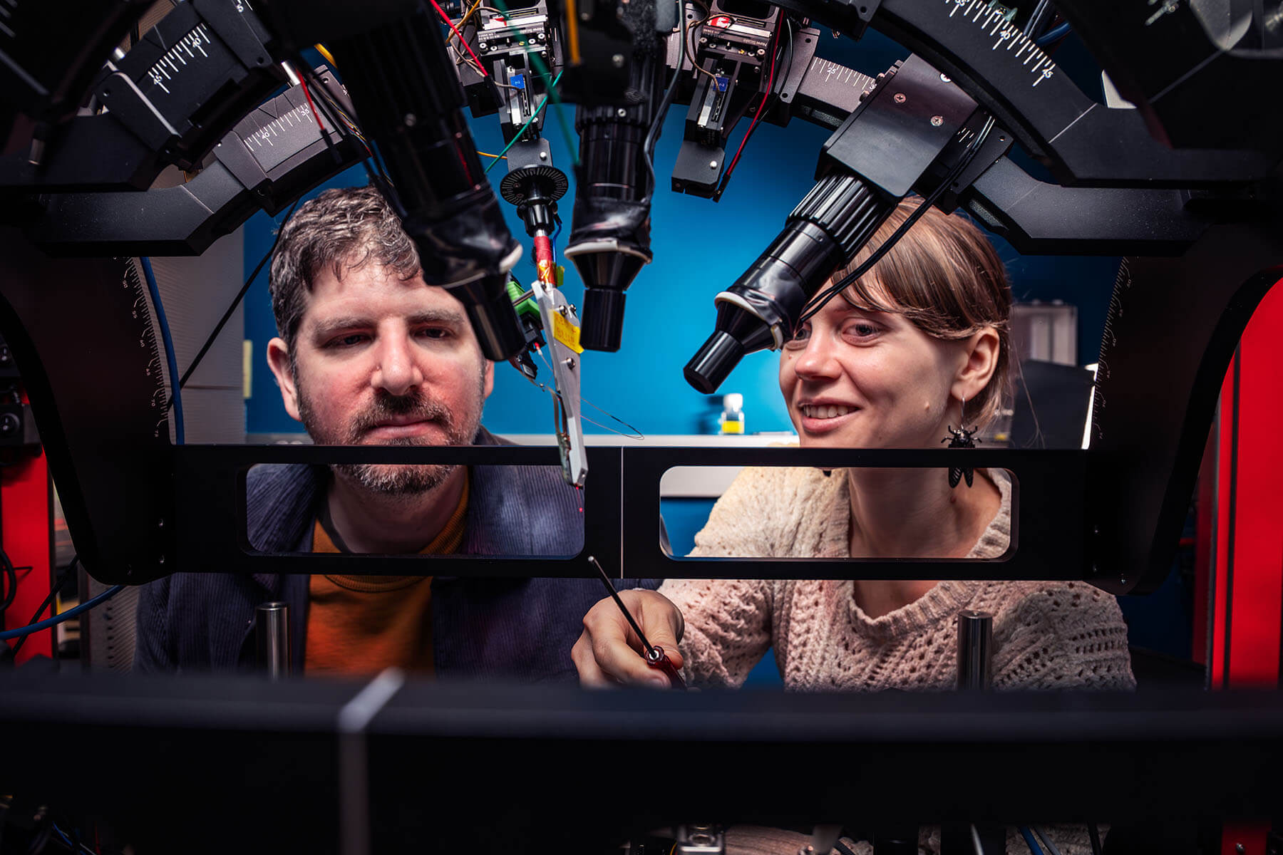
Despite its size, scientists unveil the secrets within this tiny structure daily. A team of researchers at the Allen Institute for Neural Dynamics, a division of the Allen Institute, is taking a closer look at these mini marvels by employing expansion microscopy, a technique that makes the brain three times bigger and see-through with the unlikely assistance of a chemical commonly found in disposable diapers.
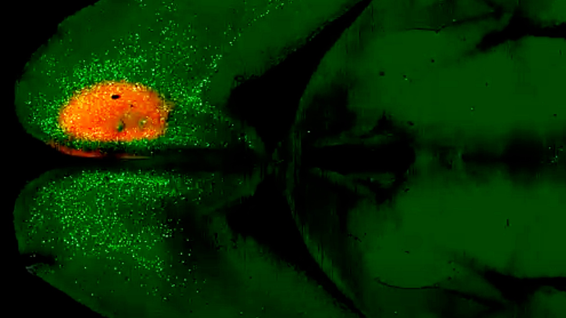
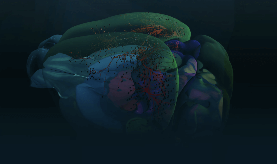
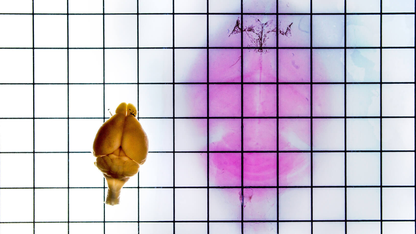
Scientists at the Massachusetts Institute of Technology first used the water absorbing chemical, known as sodium polyacrylate, to expand cells and slices of tissue. The Allen Institute team is now using such technology to expand entire mouse brains in a first-of-its-kind approach. By expanding whole mouse brains before capturing images of them, the scientists can see individual axons, the long signal-sending branches of neurons, at a much higher resolution. This is especially important for the types of neurons the Allen Institute team wants to study, which have branches that can stretch multiple centimeters in the brain but are a thousand times thinner than a human hair.
“There are really fine structures in the brain that are normally really difficult to image,” said senior research associate Naveen Ouellette. “We’ve gotten to the point where we’re now able to see these fine structures and we’re able to see them brightly labeled, which is all very difficult to do.”
In 2015, neuroscientist Ed Boyden, Ph.D., and his team at MIT shook the scientific community by using a swellable polymer to expand brain slices from a mouse to more than four times their original size. The compound contains sodium polyacrylate, which attracts moisture and expands when introduced to water. Making tissues or entire organs physically larger means scientists can capture higher levels of detail using existing microscopes. Expansion microscopy has since been used in projects ranging from neurological disorders to breast cancer biopsy analysis.
The nitty gritty of brain expansion
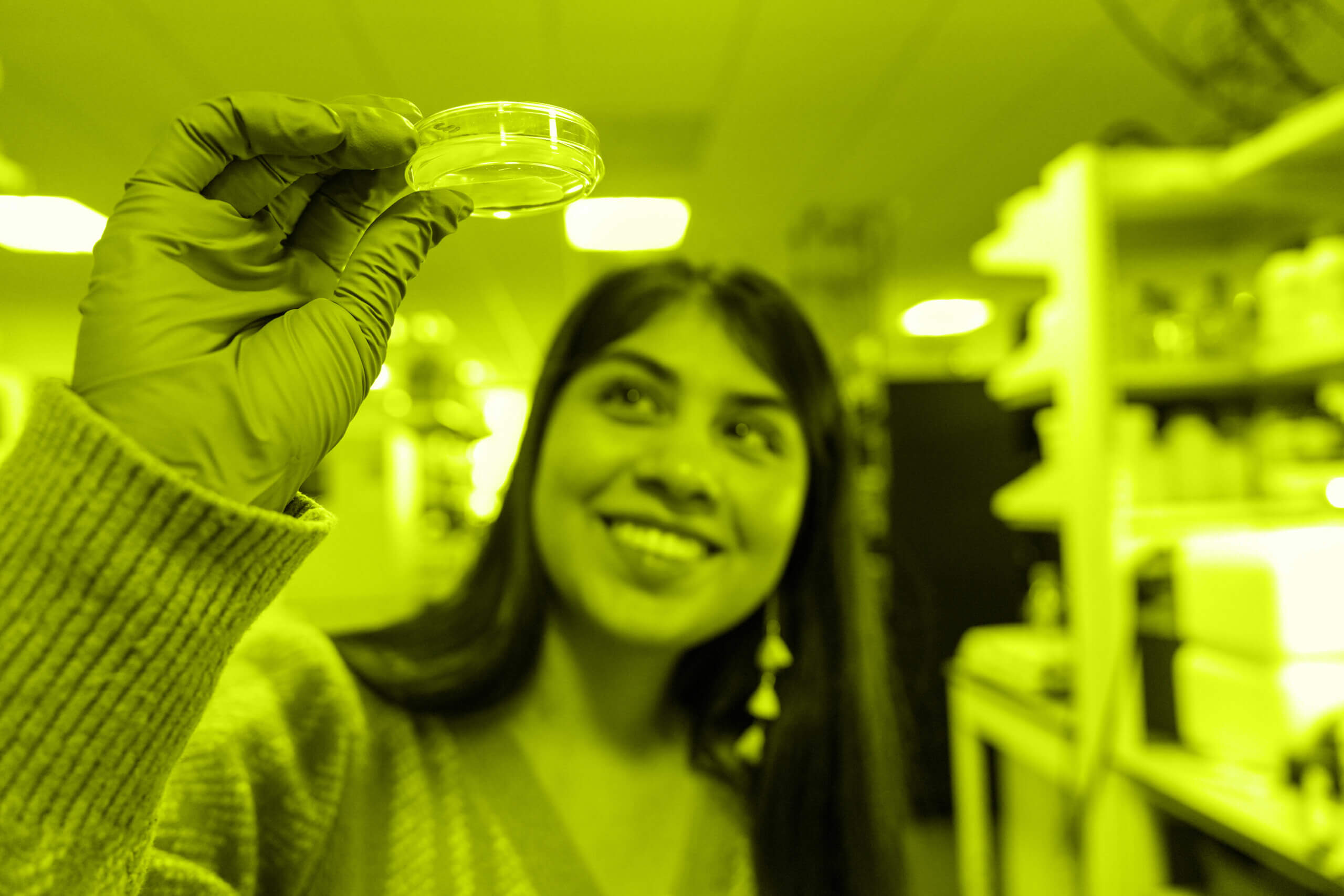
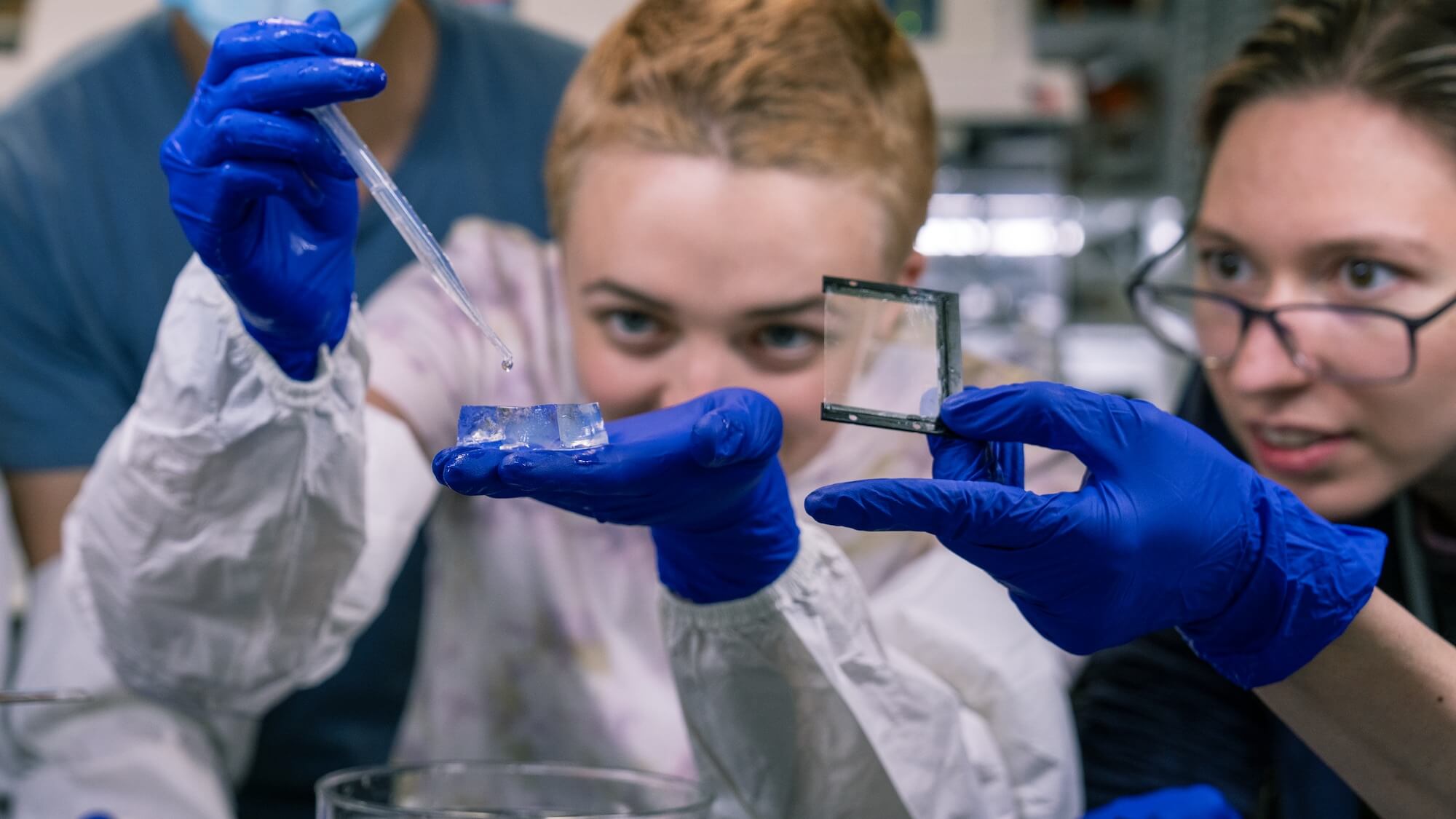
To preserve the brain’s fine spatial relationships, the Allen Institute scientists first add a molecular anchor that links the brain’s structures in proportion to each other as the whole organ is expanded. It’s akin to drawing a smiley face on a balloon before it’s blown up — the face gets bigger but maintains its same shape. In order to see the neurons they want to study, the scientists must also label specific cells with a fluorescent marker and clear the brain — rendering the organs see-through.

To get from opaque to transparent, scientists must first remove any fat in the way. These fats, or lipids, play a key role in supporting brain function. However, they block closer examination of neurons. That’s where delipidation comes in, otherwise known as de-fatting. This process uses a chemical similar to paint stripper to remove most of the fats. For that chemical to work, the scientists must first dehydrate the whole brain, which makes it even smaller. After the fats are stripped out, the brain is rehydrated and goes through a second delipidation step that includes alcohol and soap — a far more respectable choice than your typical Trader Joe’s peony foam hand soap. The entire process, including neuron labeling, delipidation, and “gelation,” can take up to two months to complete.
Getting to that point seems taxing, but creating transparent brains makes whole brain microscopy possible, allowing scientists to see the big picture of the brain at the finest detail, all at once.
“We want to image the whole brain at high resolution and a limitation that comes from traditional imaging techniques is that you can only see parts of it at a time,” said senior scientist Jayaram Chandrashekar, Ph.D. “The clarity provided by whole brain expansion overcomes this limitation. To track these individual axons, we need to be able to image the entire brain without losing any data.”

But why strive to do this anyway? It’s about understanding the anatomy of the brain at super resolution, one class of neuron at a time. Kevin Cao, another neuroscientist on the Neural Dynamics team, said: “Imagine we’re trying to map the human skeleton for the first time. This technique is like taking away all the parts of your body that aren’t your bones, and then fixing your bones to a grid and stretching them out four times bigger. They’d be in the same location relative to each other, just much bigger and their features unobstructed by tendons and muscle. In the context of the brain, we’re trying to understand how different parts of it interact with one another by mapping it in this way.”
With microscopic features, maintaining precision while increasing size is crucial. “Whole brain expansion is important because it enables us to study brain-wide connectivity at the nanoscale level,” Cao said.
Most microscopes are designed to image thin slices of a brain or other tissue; these machines provide essential information, but they can’t always capture the full breadth of neurons in the entire brain. The Neural Dynamics team also recently unveiled a new kind of microscope known as ExA-SPIM, which is purpose-built to image these giant gummy brains. Together, the equipment and the expansion technique led to an in-depth exploration of individual neurons and axons.
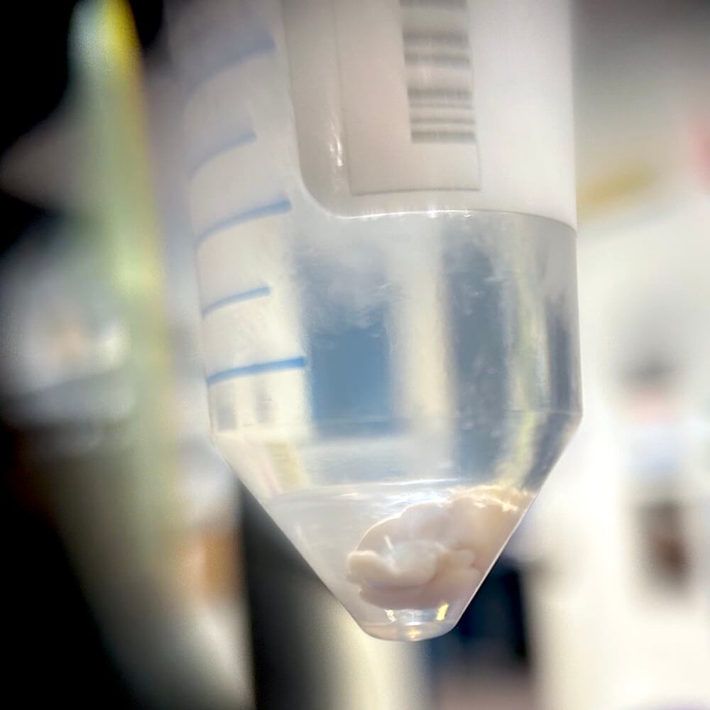
With the naked eye, it takes a certain skill level to see an expanded, cleared brain, because it is so translucent.
“It’s like a clear Jell-O shot, basically,” Ouellette said. “There’s almost like a ghost image of the brain.”
Things need to line up perfectly to even catch a glimpse. The calculated position of the wrist. The right choice of background. A subtle tilt of the neck and then an outline can be detected. It is elusive, like a black cat in the night. Thinking of the time, the effort, the possibilities, in a see-thru jelly vessel, is unexpectedly moving.
Citations
about the allen institute
The Allen Institute is an independent, 501(c)(3) nonprofit research organization founded by philanthropist and visionary, the late Paul G. Allen. The Allen Institute is dedicated to answering some of the biggest questions in bioscience and accelerating research worldwide. The Institute is a recognized leader in large-scale research with a commitment to an open science model. For more information, visit alleninstitute.org.