in this article
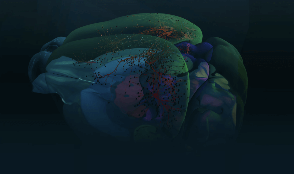
A global consortium of scientists has created the first and most detailed “developmental maps” of the mammalian brain (from mouse to human) to date, taking the first critical steps in unraveling the mystery of early brain development and the vital role it plays in health and disease.
Life-altering neurodevelopmental disorders that lead to significant cognitive, communication, behavioral, or psychomotor impairments affect 15% of children or adolescents worldwide, with diagnoses of autism and attention deficit hyperactivity disorders (ADHD) increasing in the United States. In humans, this early phase of brain development is uniquely long, so understanding this critical phase where things can go wrong is essential for illuminating our understanding of and treating brain disorders.
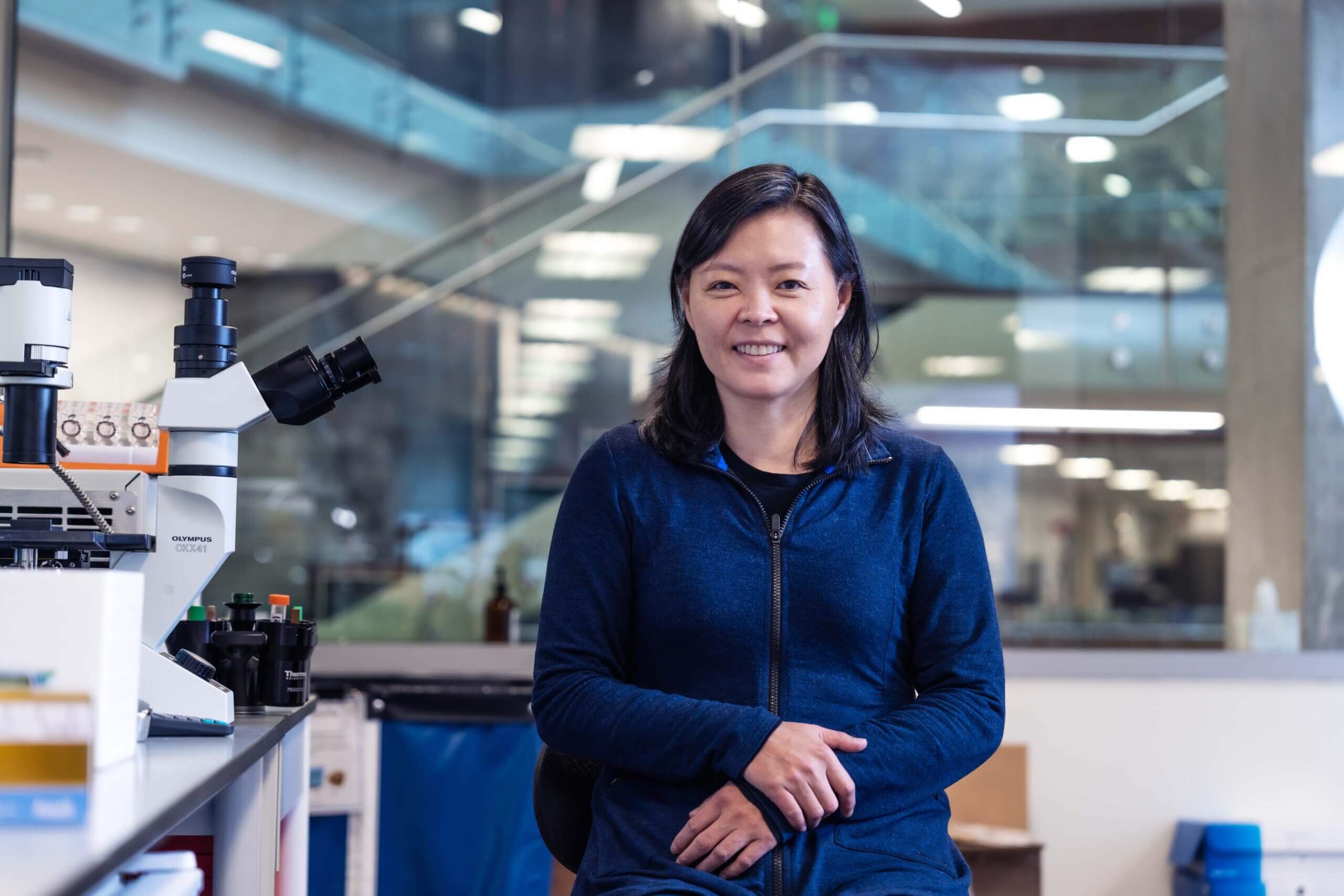
“This set of work gives us a detailed blueprint of how different brain cell types emerge and mature over time,” said Hongkui Zeng, Ph.D., executive vice president and director of Brain Science at the Allen Institute and one of the lead authors on several of the studies.
By understanding when and where critical genes are turned on during development, we can begin to uncover how disruptions in that process may lead to disorders like autism or schizophrenia. It’s foundational knowledge that opens the door to better diagnoses and targeted treatments.
Hongkui Zeng, Ph.D, executive vice president and director of Brain Science at the Allen Institute
In a package of 12 studies published in the Nature family of journals, researchers reveal new features of mammalian cell types during early development and begin to shed light on the environmental factors—including sensory inputs and social behavior—that affect how brains develop. The resulting comprehensive, cross-species developing brain cell atlases lay the foundation and provide powerful tools for a new era in the understanding of the human brain in health and disease.
“It’s really been a long-standing mystery to understand how these processes occur,” said Tomasz Nowakowski, Ph.D., one of the study authors and associate professor of neurological surgery, anatomy, psychiatry and behavioral sciences at the University of California, San Francisco. “Building on the findings from the adult brain and venturing into the developmental stages is profoundly important because it is going to inform our understanding of the vulnerabilities and mutations which can lead to neurodevelopmental disorders.”
This landmark achievement was supported by the National Institutes of Health’s Brain Research Through Advancing Innovative Neurotechnologies® Initiative, or The BRAIN Initiative®, aimed at accelerating the development of innovative neurotechnologies and revolutionizing our understanding of the human brain.
“These maps are a phenomenal achievement, and very important references that are sorely needed as we seek to understand the mechanisms governing the developing brain in health and disease,” said Joshua Gordon, M.D./Ph.D., former director of the National Institute of Mental Health and current chair of Columbia University’s Department of Psychiatry. “They provide a scaffold on which we can construct a deeper understanding of autism, schizophrenia and other disorders that we know unfold during brain development.”
Allen Institute scientists contributed three studies to this package. Transcriptomic and spatial organization of telencephalic GABAergic neurons focuses on a special group of brain cells in mice called GABAergic inhibitory neurons. These cells act like brakes in the brain, calming down excessive activity and helping different brain regions communicate smoothly. These neurons in humans are critical for everything we do, from movement and memory to managing emotions.
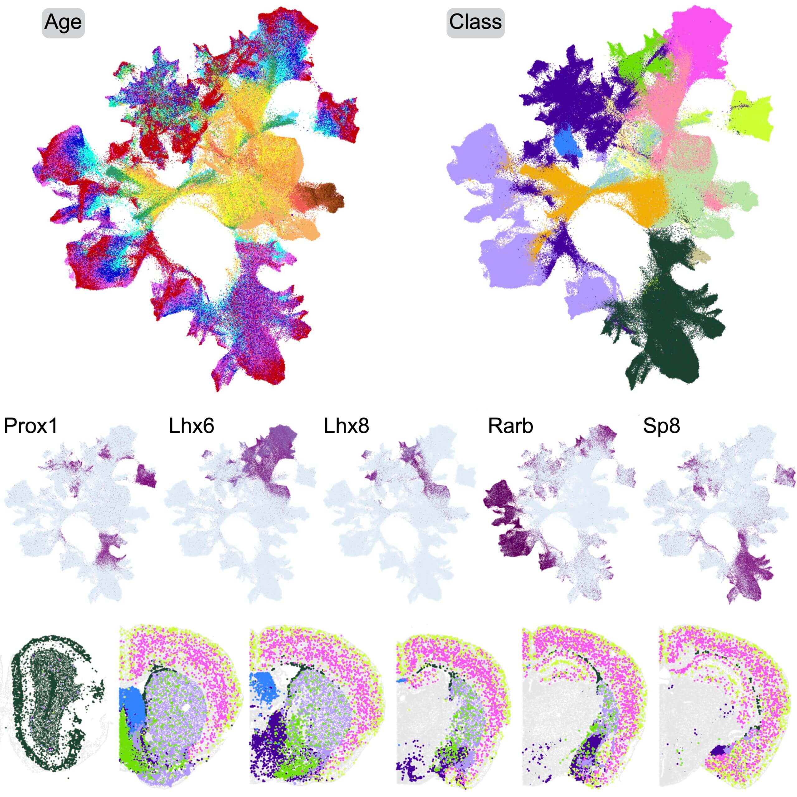
Using data from over 1.2 million brain cells, the researchers created the most complete “family tree” of these cells to date, showing how they develop, spread out, and differentiate into distinct subtypes. One of the most striking findings shows how these cells travel long distances from where they are born to where they end up—sometimes crossing entire brain regions. Scientists also discovered that some of these neurons keep developing long after birth, especially in parts of the brain involved in learning, decision making, and emotions. This means there may be a longer window than previously thought for intervening and helping the brain rewire itself, particularly for children with developmental challenges. Researchers can now also identify which specific subtypes of GABAergic neurons are affected in different disorders and develop more targeted therapeutic approaches.
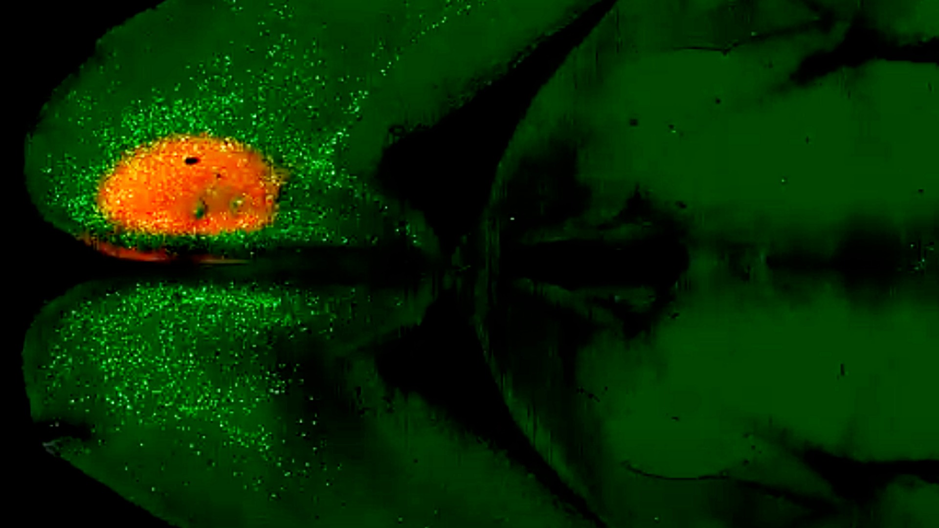
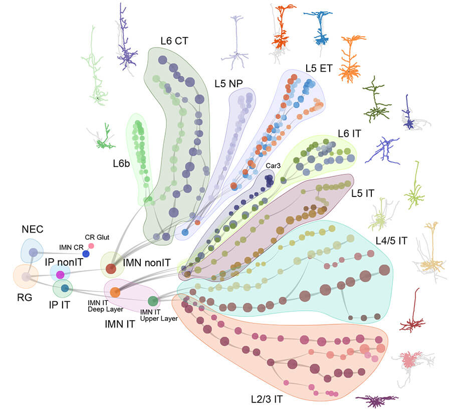
In a second study, Continuous cell type diversification in mouse visual cortex development, scientists turned their attention to the visual cortex, the part of the brain that processes what we see. They tracked over 770,000 individual cells from the earliest days of brain development through young adulthood in mice and constructed developmental trajectory trees for all the cell types in the visual cortex. They learned that brain cells don’t finish developing before birth like once thought. Instead, new cell types continue to form well into the animal’s youth, especially around key moments like when its eyes first open or when the brain starts learning to process visual information. This finding suggests that experiences after birth—like seeing, hearing, or interacting with the world—may shape brain development far more than we realized. It also means that developmental disorders might still be treatable after birth during these “critical periods” when the brain is still building and refining its circuits.
In Whole-cortex in situ sequencing reveals input-dependent area identity, researchers use BARseq, a highly specialized genetic sequencing technique, to map the gene expression of millions of single neurons across the cerebral cortex. Researchers discovered that the unique combination of different neuron types acts like a “cellular signature” that predicts and defines the brain region. A crucial finding was that sensory experience, such as vision, is also strongly linked with brain region specialization during development. Together, these studies reveal the exact timing and patterns by which brain cells grow, specialize, and connect. These discoveries reveal critical windows during development—some even after birth—when the brain is especially sensitive to change. This valuable insight has implications for understanding and treating childhood brain disorders that begin in life’s earliest stages.
Citations
about the allen institute
The Allen Institute is an independent, 501(c)(3) nonprofit research organization founded by philanthropist and visionary, the late Paul G. Allen. The Allen Institute is dedicated to answering some of the biggest questions in bioscience and accelerating research worldwide. The Institute is a recognized leader in large-scale research with a commitment to an open science model. For more information, visit alleninstitute.org.