in this article
Looking at living neurons from one region of the cortex, the outermost part of the mammalian brain that is responsible for our higher cognitive functions, the research team found that one kind of human neuron sends and receives electrical signals in a different way than does the mouse version of the same cell. The researchers published a study in print today in the journal Neuron describing their comparison.
The researchers found that the human neurons are studded with groups of proteins known as h-channels, while the mouse cells of this class are largely devoid of h-channels.
That finding could have implications for mouse models of human brain disorders and for drug development studies, the researchers said. If drugs are developed that act on h-channels, they’d likely have different effects in humans than they would in mouse studies, which are often a necessary precursor to clinical trials.
“If human neurons have different electrical properties than rodent neurons of the same type, it has implications for how you identify dysfunctions relevant to a brain disorder, or even how you’d test a new therapeutic and how it works,” said Jonathan Ting, Ph.D., a neuroscientist at the Allen Institute for Brain Science, a division of the Allen Institute, and senior author on the study.
That’s not just a hypothetical concern. An existing epilepsy drug, lamotrigine, acts in part by changing h-channel activity. The research team hasn’t yet shown that the human and mouse neurons respond differently to different drugs — that’s on their future to-do list. But because h-channels act as a gatekeeper between the neuron and its environment, letting in or out certain atoms during brain signaling in response to changes in voltage across the cell surface, it’s reasonable to assume these channels could affect how the brain responds to some drugs which would also act from the outside of the cell.
A different wavelength of brain signal
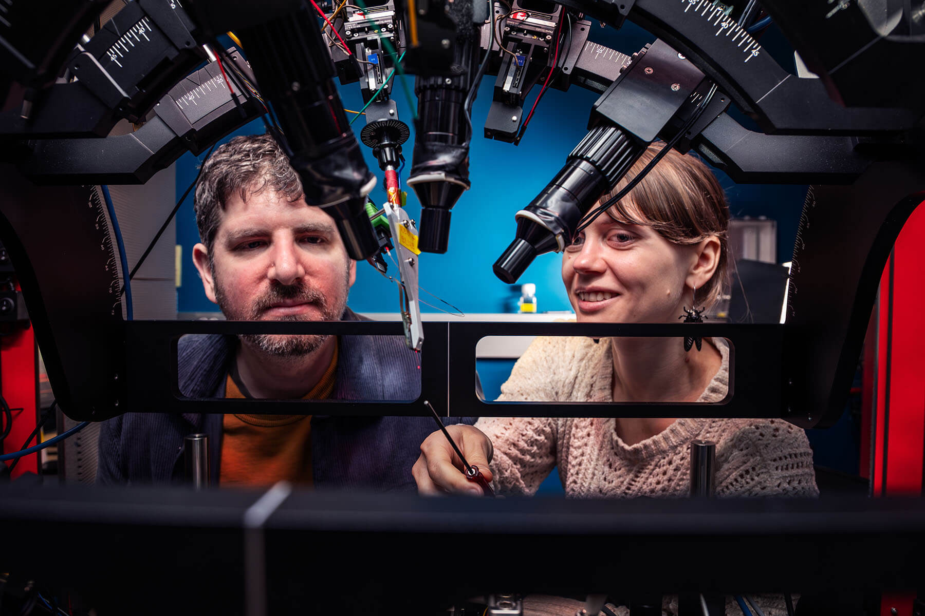
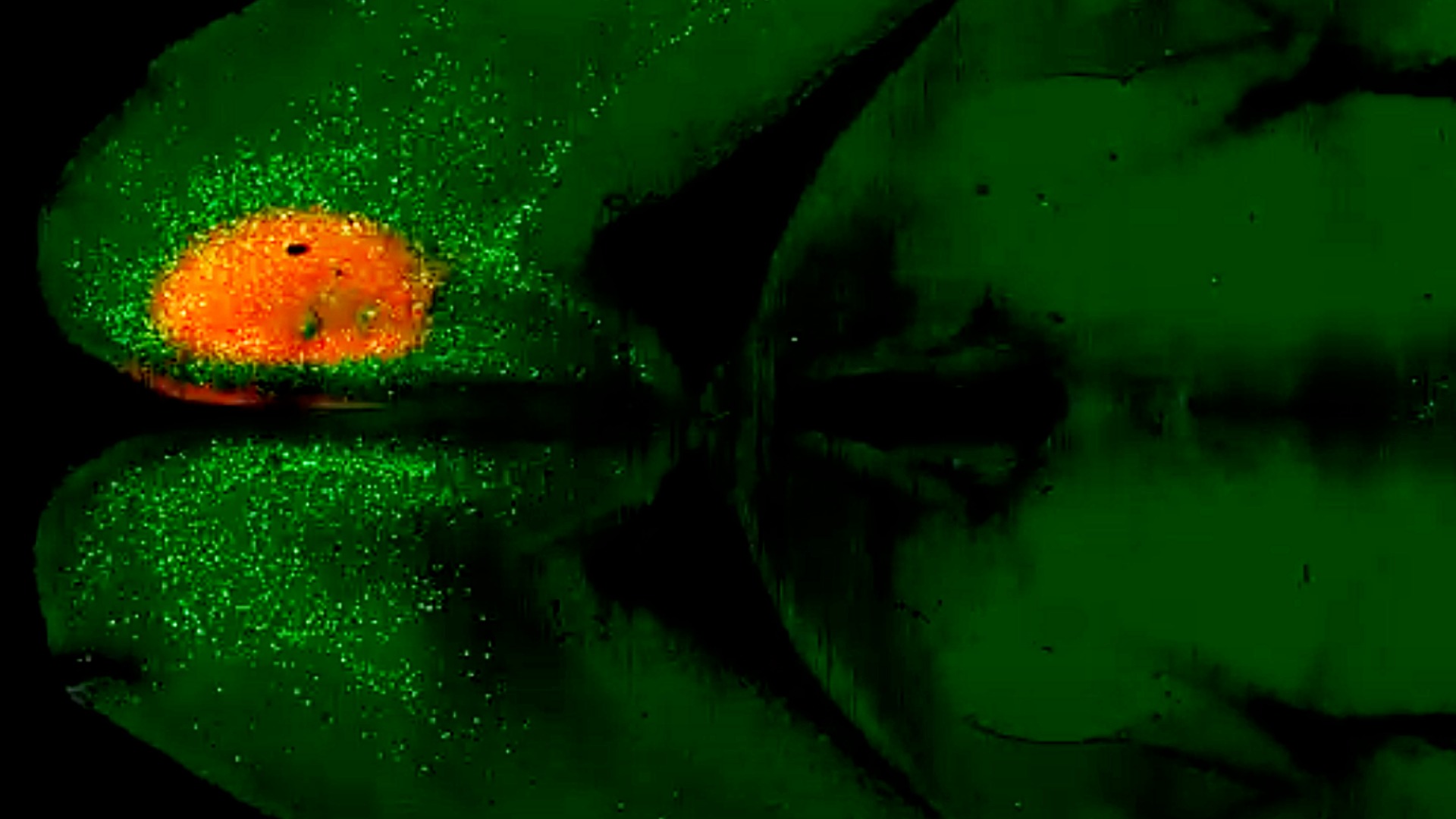
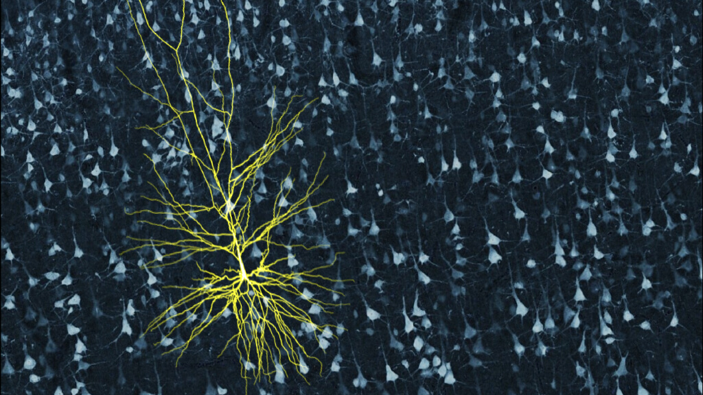
To look at the live human cells, which are known as pyramidal neurons, the researchers used small pieces of human brain that were removed during surgery by neurosurgeons at UW Medicine, Harborview Medical Center and the Swedish Neuroscience Institute. Patients who undergo surgery for epilepsy or brain tumor and who consent to be part of the Allen Institute research donate these tiny bits of their brain that have to be removed for the surgeons to reach the diseased areas.
At first glance, the human pyramidal neurons from this particular brain region, the second and third layer of the six-layered cortex, might not appear very different from their mouse counterparts. The human versions are clearly larger, but they have a similar structure to the mouse neurons. But when the scientists started taking readings from the live cells to measure the electrical signals these cells send and receive, they saw immediate variations between mouse and human.
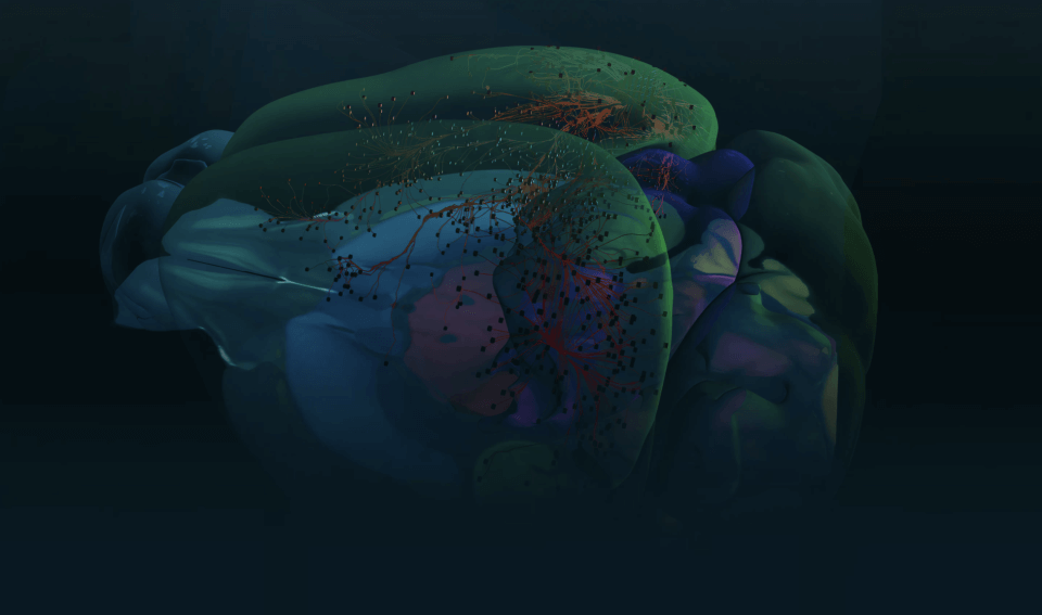
To understand what those signals mean, the research team turned to computational modeling. The models revealed that h-channels likely tune the human cells to a different wavelength of electrical signal. The model predicted that the human neuron responds best to brain waves in the theta frequency, a slower wavelength that’s been implicated in learning, memory and attention.
“You could think of the h-channels like a transistor,” said Costas Anastassiou, Ph.D., a computational neuroscientist at the Allen Institute for Brain Science who led the modeling part of the study. “Depending on the hardware, it will affect the abilities of that circuit.”
That difference in hardware could even relate to the differences in cognitive functions between mice and humans (or between mice and primates in general).
“In terms of the frequency of signal they prefer, these cells are totally different from what we see in the mouse cell,” said Brian Kalmbach, Ph.D., a neuroscientist at the Allen Institute for Brain Science and first author on the paper. “These sorts of frequencies are associated with attention and learning and memory, so we might speculate that since these cells are tuned into this sort of input, they’re participating in functions like those.”
Surgical limitations
There’s a caveat to the h-channels study — the live human neurons were derived from patients with brain diseases, either epilepsy or brain tumors. There’s really no other way researchers can study live human neurons other than through these surgical collaborations and with the assistance of patients who agree to donate their tissue, and only serious diseases necessitate brain surgery.
“It’s very cool to be involved in this work — this is really cutting-edge stuff — but we have to remember these conclusions are being drawn from non-normal brains,” said Daniel Silbergeld, M.D., a neurosurgeon at UW Medicine who specializes in brain tumors and who collaborated on the study.
Could the increased h-channels and the resulting change in signaling just be a property of diseased brains? The researchers don’t think so.
“There are a few lines of evidence that lead us to believe this phenomenon is not solely due to disease,” Kalmbach said.
For one, they see the same properties in brain tissue excised from patients with brain cancer or epilepsy, two very different diseases. This tissue is coming from parts of the brain that are far from the tumor or central focus of seizures. The researchers also saw evidence for high levels of h-channel gene activity in the same cells in non-diseased postmortem human brain tissue, although they can’t test the signaling properties of dead cells.
Next steps
To obtain deeper insights into what this protein is doing in these human neurons and where the differences arose in the evolutionary path between mice and people, the researchers need to study more species – ideally, species that are closer cousins to humans than mice are, such as monkeys. As in humans, the region of the cortex the researchers studied is also much larger in monkeys than it is in mice.
“Layer 3 is the layer of cortex that has really expanded in humans over other mammals,” Silbergeld said. “This study is kind of a step toward defining what this expanded layer of neurons does and how it’s changed from other mammals.”
Other authors on the study are Anatoly Buchin, Brian Long, Jennie Close, Anirban Nandi, Jeremy A. Miller, Trygve E. Bakken, Rebecca D. Hodge, Peter Chong, Rebecca de Frates, Kael Dai, Zoe Maltzer, Rusty Nicovich, C. Dirk Keene, Christof Koch and Ed Lein of the Allen Institute for Brain Science; Ryder Gwinn and Charles Cobbs of Swedish Neuroscience Institute; and Andrew Ko and Jeffrey Ojemann of the University of Washington School of Medicine.
Citations
about the allen institute
The Allen Institute is an independent, 501(c)(3) nonprofit research organization founded by philanthropist and visionary, the late Paul G. Allen. The Allen Institute is dedicated to answering some of the biggest questions in bioscience and accelerating research worldwide. The Institute is a recognized leader in large-scale research with a commitment to an open science model. For more information, visit alleninstitute.org.