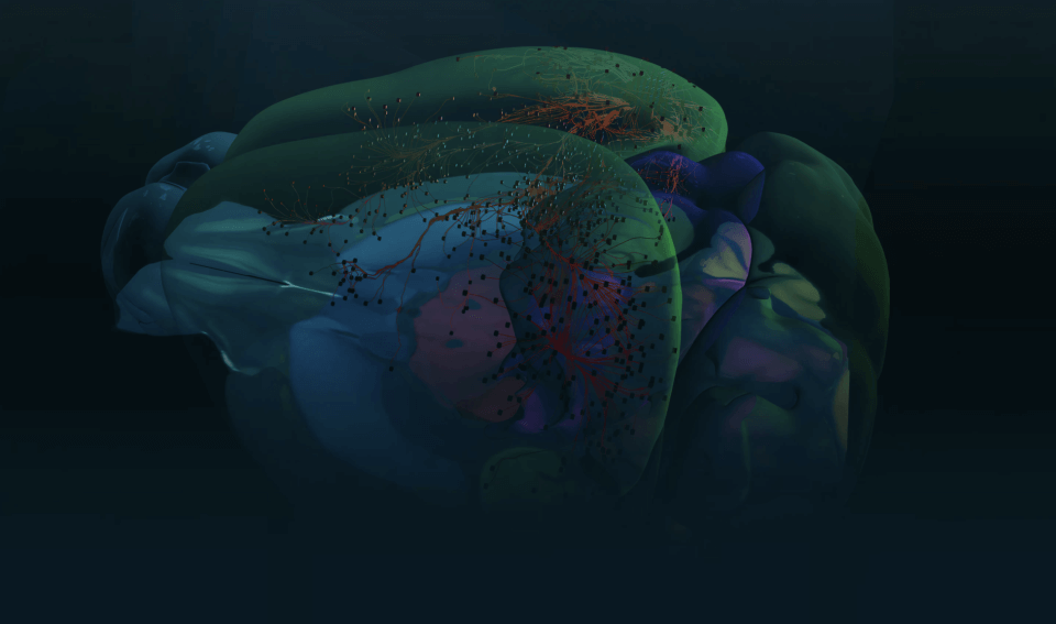

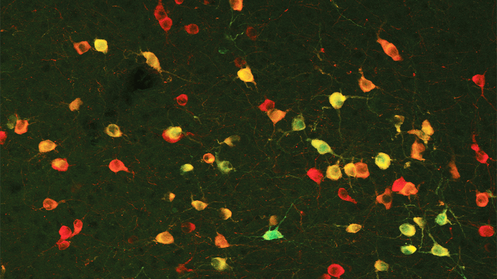
From shell to stem, our brains are chock full of neurons. They chatter incessantly so we can accomplish everything from breathing to reading this article.
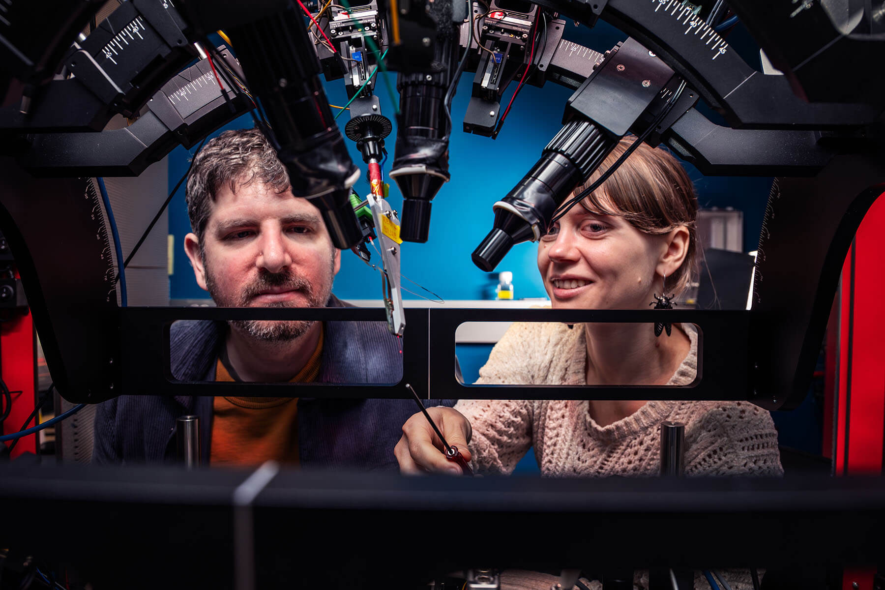
In order to understand how our brains work, neuroscientists at the Allen Institute for Brain Science, a division of the Allen Institute, research some of its most fundamental units: the neurons. By studying attributes such as neurons’ shapes, electrical signals and the genes they turn on and off, neuroscientists come one step closer to understanding what they do.
Neurons use their unique characteristics to act in one of two broad ways: to excite or inhibit. Excitatory neurons activate other neurons, while inhibitory neurons suppress other neurons’ activity. Researchers study how these neurons work in complex circuits to form our brains’ cohesive emotions, actions and reactions.
We asked five neuroscientists at the Allen Institute for Brain Science to describe their favorite neuron.
‘The life of the party’: The ET neuron
There’s a specific group of excitatory neurons that Allen Institute researchers who study the human brain are particularly interested in, known as extratelecephalic-projecting neurons, or ET neurons. This class of neurons includes the rare von Economo neuron, which is found only in humans and a handful of other mammals. ET neurons sit in the outermost shell of our brain, the cortex, and convey information to deeper brain areas. The cortex is made up of upper and lower layers. In the human brain, ET neurons are only found in the lowest layers, and their axons extend out to communicate with even deeper brain areas.
“Anything that the cortex does to affect our behavior is routed through these cells,” said Allen Institute neuroscientist Brian Kalmbach, Ph.D., who studies how ET neurons send and receive electrical signals in the human brain.
You can think of ET neurons as “the life of the party,” he said. They’re big, boisterous and talking to everyone.
Kalmbach records from ET neurons as they chatter away to other neurons in live human brain tissue, which is donated from patients undergoing brain surgery for epilepsy or tumors.
The Shh-ing chaperone: The fast-spiking interneuron
Fast-spiking interneurons, a type of inhibitory neuron, are sprinkled throughout the human cortex to prevent hyperactivity in excitatory neurons. That’s an essential job, because too much activity in the brain can lead to seizures.
“They’re like a chaperone for a kindergarten field trip,” said neuroscientist John Mich, Ph.D. “There is one adult for every 10 kids, and the chaperone is just trying to keep them in line and make sure they do their job well.”
In this analogy, the kids are the excitatory neurons. The chaperones, the fast-spiking interneurons, can spring into action 10 times faster than the neurons they connect with, allowing them to prevent seizures.
“They’re adamant about saying ‘shh, everybody calm down,’” Mich said.
Mich and his colleagues are harnessing the power of these shh-ing cells to research new therapies for Dravet Syndrome, an uncommon but severe form of early-childhood epilepsy.
Dravet Syndrome is caused by a single mutation in a gene that is responsible for the interneurons’ ability to quickly switch on. In children with the disease-causing mutation, the interneurons cannot activate fast enough to quiet hyperactivity in the brain, leading to the disease’s hallmark seizures.
Mich and his colleagues are working on a new kind of gene therapy to deliver non-mutated copies of the gene directly to fast-spiking interneurons in the hopes of restoring their ability to prevent seizures in people with Dravet Syndrome.
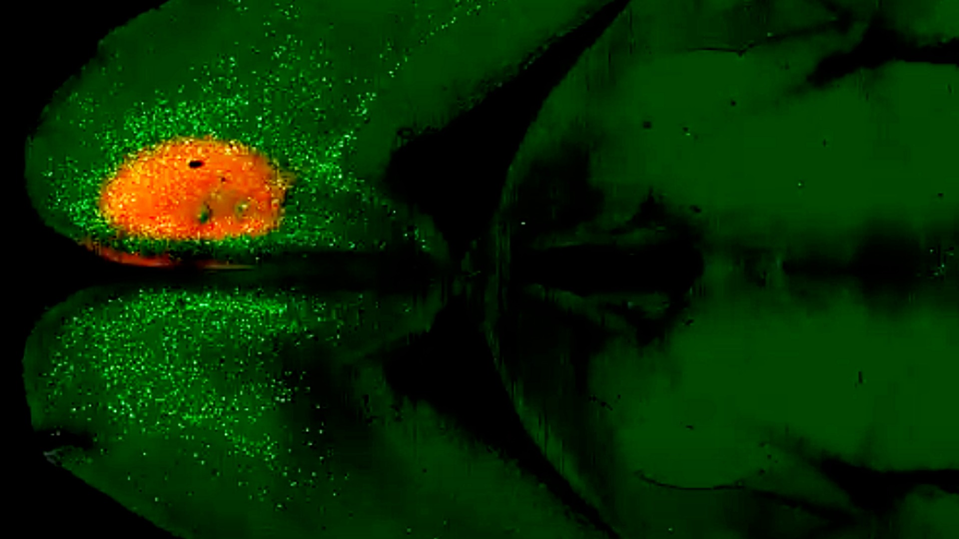
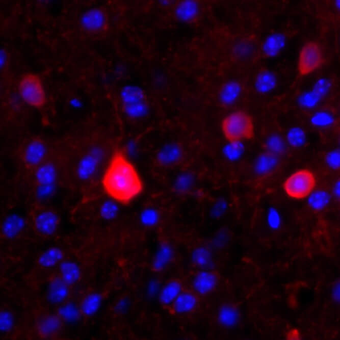
A rare gem: The cholinergic neuron
Cholinergic neurons are rare in the human brain. A handful of these inhibitory neurons sit in our cortex, right behind our foreheads.
Cholinergic neurons have axons that branch from their bodies like a starburst, a characteristic shape that allows them to reach many other cells in the brain despite their scarcity. These rare neurons use a molecule known as acetylcholine to convey information about movement, cognition and body temperature.
“I think it’s a proxy for life, how one person can have a big impact. One little cell can have a very big impact on overall function,” said Allen Institute neuroscientist Tanya Daigle, Ph.D.
When cholinergic neurons don’t work as they should, the cortex loses its ability to talk to other parts of the brain. Dysfunction of these rare cells has been seen in brain disorders including Alzheimer’s Disease, Parkinson’s Disease and ADHD.
Before coming to the Allen Institute, Daigle researched drug therapies that improved cholinergic neuron function with the hopes of treating both Alzheimer’s and Parkinson’s Diseases. These drugs are currently on the market.
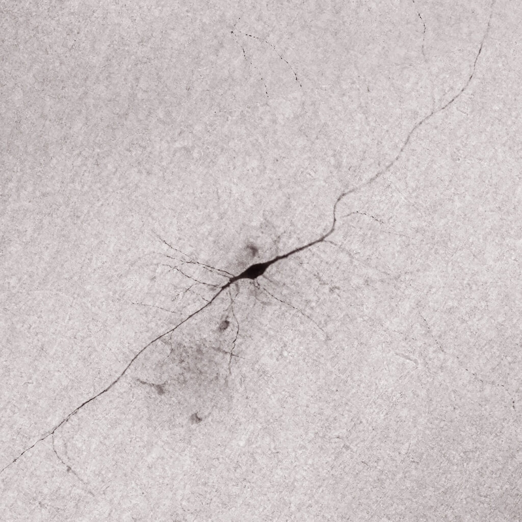
New kid on the block: The SST met-type 7 neuron
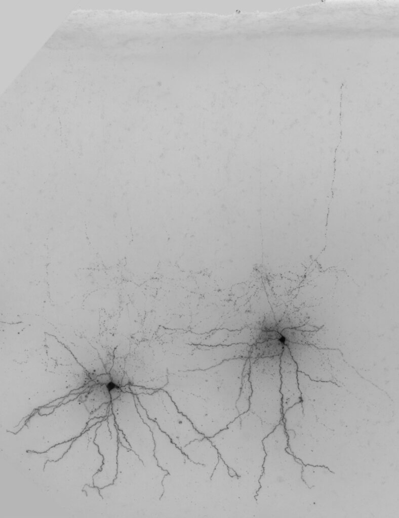
Researchers at the Allen Institute group neurons into families to study their shared traits. Through this work, neuroscientist Staci Sorensen, Ph.D. was recently introduced to her favorite neurons, the newest members of a family of inhibitory neurons: the SST met-type 7 neurons. Sorensen and her colleagues at the Allen Institute are experts in neurons’ detailed 3D shape. In their work characterizing a family of mouse inhibitory neurons, the SST neurons, they found that SST met-type 7 neurons look very different from their close cousins.
“There are all these SST types in the lower layers of cortex that have not been characterized or appreciated at all,” Sorensen said.
SST met-type 7 neurons are shaped like spiders descending from a web. They sit in the lower layers of the mouse cortex, transitioning from the brain’s shell to its core. While other neurons in the SST family predominantly send their axons to the surface of the cortex, these newly discovered neurons contain most of their axons in the lower layers, and apart from one or two branches, stop abruptly at the lower layer border.
Researchers don’t yet know what these neurons do in the brain, but Sorensen has a prediction: SST met-type 7 neurons could be quieting the excess noise of nearby excitatory neurons. This would in turn enable a more precise transfer of information from the cortex to other brain areas.
Shh-ing the shh-ers: The VIP neuron
Excitatory neurons receive mixed messages. They are bombarded by different inhibitory neurons whispering, “Shh, don’t say that, say this.” Luckily, excitatory neurons have a trusty friend who helps them decide whom to listen to: the VIP neuron. VIP neurons quiet other inhibitory neurons, freeing the excitatory neurons to send their signals under certain circumstances.
“They have a net positive effect on excitatory cells even though they’re an inhibitory class,” neuroscientist Marina Garrett, Ph.D., said of her favorite neurons, the VIP neurons.
Garrett is working with other behavioral neuroscientists at the Allen Institute to determine why VIP neurons work the way they do. She has a prediction.
“They’re going and telling everybody else what’s salient, what’s relevant to pay attention to,” Garrett said.
Sometimes, the relevant information is timing, like quitting time at the end of a workday or the time the bus comes. Other times, the relevant information is novelty, like meeting a new person or discovering a piece of cake in the fridge. Either way, VIP neurons might help excitatory neurons decide what to pay attention to.
“There’s a ton we still don’t know,” Garrett said. “We have a lot of suggestive information, but putting it into a true mechanistic understanding requires a lot more data, better tools, and the ability to dissect the circuit in new ways.”
Citations
about the allen institute
The Allen Institute is an independent, 501(c)(3) nonprofit research organization founded by philanthropist and visionary, the late Paul G. Allen. The Allen Institute is dedicated to answering some of the biggest questions in bioscience and accelerating research worldwide. The Institute is a recognized leader in large-scale research with a commitment to an open science model. For more information, visit alleninstitute.org.